The GFAP Monoclonal Antibody (CAB19058) is a high-quality antibody developed for reliable detection and analysis of target proteins. Astrocytes play a significant role in supporting neuronal function and maintaining the blood-brain barrier, making GFAP a valuable marker for studying neuroinflammation, brain injury, and neurodegenerative diseases.This antibody, generated in rabbits, demonstrates high reactivity with human samples and is validated for use in various applications, including immunohistochemistry and immunofluorescence.
This antibody is validated for use in WB, IHC-P, ELISA, IF-F, IF-P, mIHC applications and has demonstrated reactivity against Human, Mouse, Rat samples.
Product Name:
GFAP Monoclonal Antibody
SKU:
CAB19058
Size:
20μL, 100μL
Reactivity:
Human, Mouse, Rat
Clone Number:
ARC0206
Conjugate:
Unconjugated
Immunogen:
A synthetic peptide corresponding to a sequence within amino acids 1-100 of human GFAP (P14136).
Recommended starting concentration is 1 μg/mL. Please optimize the concentration based on your specific assay requirements.
Synonyms:
ALXDRD, GFAP
Positive Sample:
U-251 MG, Mouse brain, Rat brain
Cellular Localization:
Cytoplasm.
Calculated MW:
50kDa
Observed MW:
50kDa
This gene encodes one of the major intermediate filament proteins of mature astrocytes. It is used as a marker to distinguish astrocytes from other glial cells during development. Mutations in this gene cause Alexander disease, a rare disorder of astrocytes in the central nervous system. Alternative splicing results in multiple transcript variants encoding distinct isoforms.
Purification Method
Affinity purification
Gene ID
2670
RRID
AB_2862551
Buffer Information
Store at -20℃. Avoid freeze / thaw cycles. Buffer: PBS with 0.09% Sodium azide,0.05% BSA,50% glycerol,pH7.3.
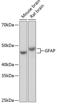
Western blot analysis of various lysates using GFAP Rabbit mAb (CAB19058) at 1:1000 dilution incubated overnight at 4℃. Secondary antibody: HRP-conjugated Goat anti-Rabbit IgG (H+L) (CABS014) at 1:10000 dilution. Lysates/proteins: 25μg per lane. Blocking buffer: 3% nonfat dry milk in TBST. Detection: ECL Basic Kit (AbGn00020). Exposure time: 1s.
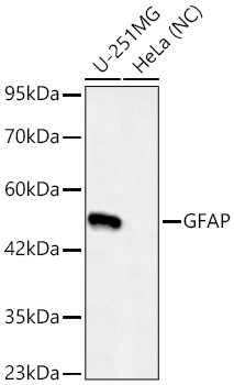
Western blot analysis of various lysates using GFAP Rabbit mAb (CAB19058)at 1:1000 dilution incubated overnight at 4℃. Secondary antibody: HRP-conjugated Goat anti-Rabbit IgG (H+L) (CABS014) at 1:10000 dilution. Lysates/proteins: 25 μg per lane. Blocking buffer: 3% nonfat dry milk in TBST. Detection: ECL Basic Kit (AbGn00020). Negative control (NC): HeLa Exposure time: 45s.
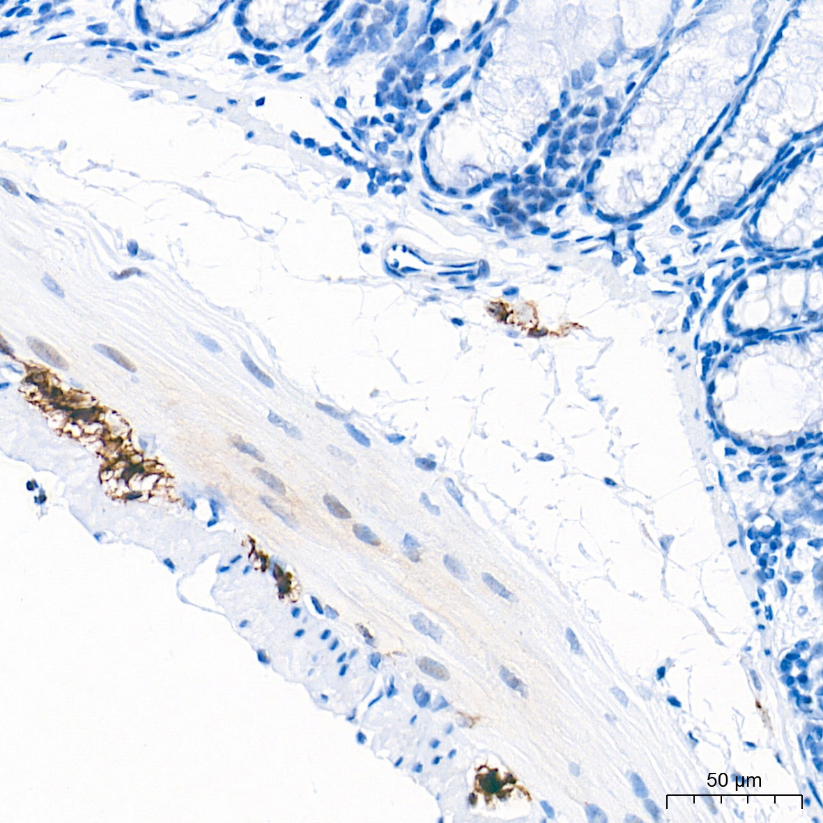
Immunohistochemistry analysis of paraffin-embedded Mouse colon tissue using GFAP Rabbit mAb (CAB19058) at a dilution of 1:500 (40x lens). High pressure antigen retrieval performed with 0.01M Tris-EDTA Buffer (pH 9.0) prior to IHC staining.
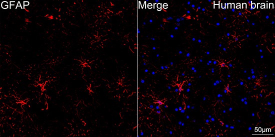
Confocal imaging of paraffin-embedded Human brain tissue using GFAP Rabbit mAb (CAB19058, dilution 1:1000) followed by a further incubation with Cy3 Goat Anti-Rabbit IgG (H+L) (CABS007, dilution 1:500) (Red). DAPI was used for nuclear staining (Blue). High pressure antigen retrieval performed with 0.01M Citrate Buffer (pH 6.0) prior to IF staining. Objective: 40x.
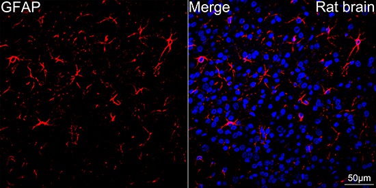
Confocal imaging of paraffin-embedded Rat brain tissue using GFAP Rabbit mAb (CAB19058, dilution 1:2000) followed by a further incubation with Cy3 Goat Anti-Rabbit IgG (H+L) (CABS007, dilution 1:500) (Red). DAPI was used for nuclear staining (Blue). High pressure antigen retrieval performed with 0.01M Citrate Buffer (pH 6.0) prior to IF staining. Objective: 40x.
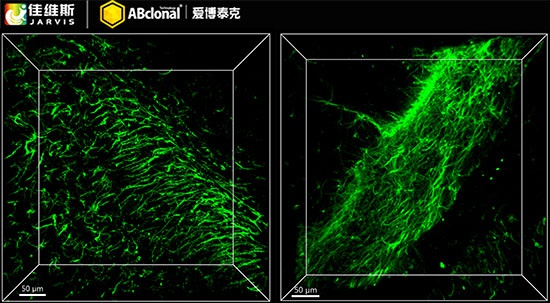
3D imaging of solvent-cleared mouse brain sections (at a thickness of 1 mm) using GFAP Rabbit mAb (CAB19058, diluted at a ratio of 1:200) . FDISCO JA11011 was used for sample clearing. We acknowledge Javis (Wuhan) Bio - Pharma Co., Ltd. in 3D imaging processing and kindly providing this image.
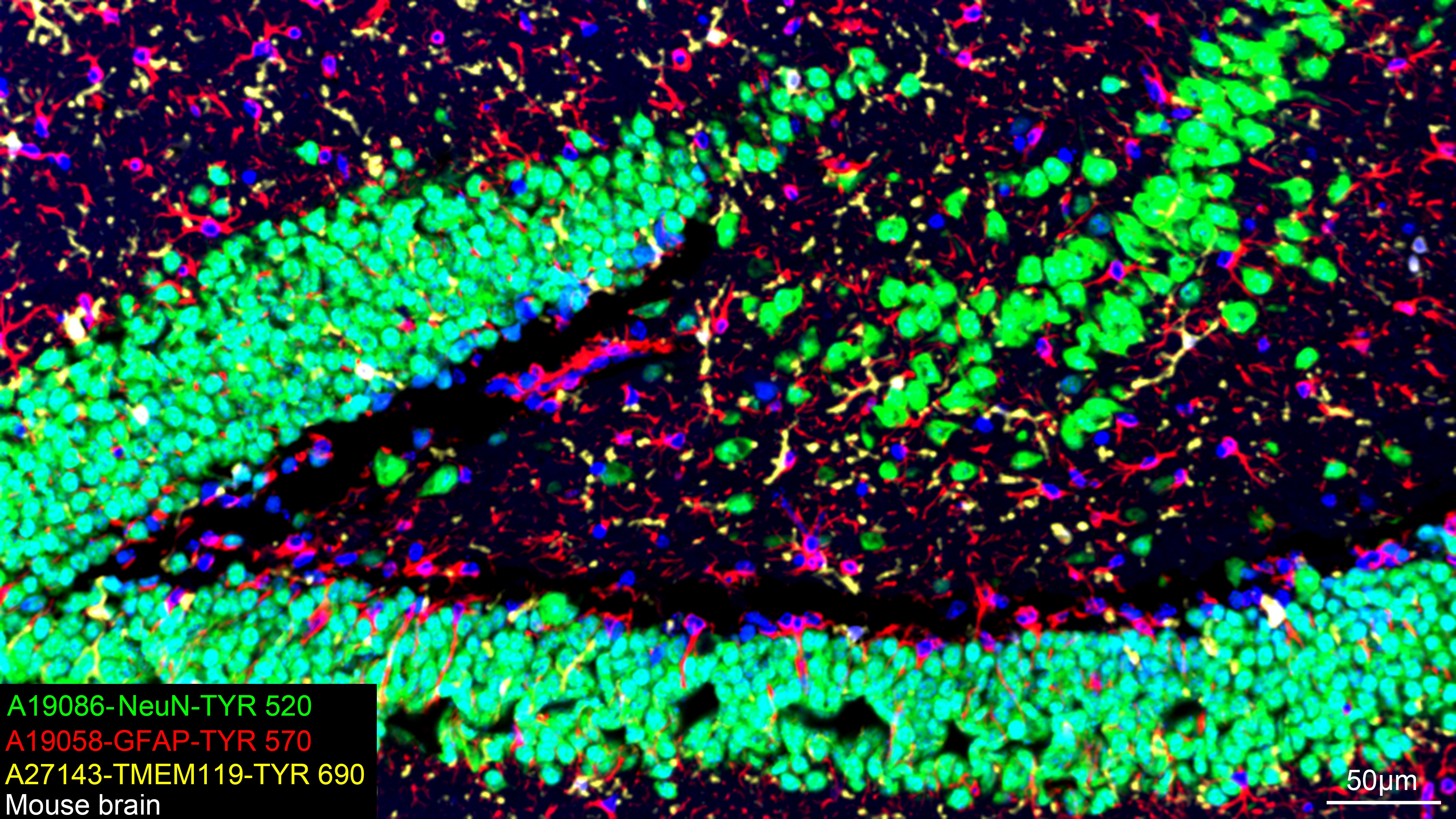
The multiplex IHC analysis on paraffin-embedded Mouse brain tissue using the following specific primary antibodies and tyramide signal amplification (TSA) reagents (RK05903) : NeuN Rabbit mAb (CAB19086, 1:2000) with TSA-TYR-520 (Green), GFAP Rabbit mAb (CAB19058, 1:500) with TSA-TYR-570 (Red), and TMEM119 Rabbit mAb (CAB27143, 1:600) with TSA-TYR-690 (Yellow). DAPI (Blue) was used for nuclear staining. Prior to multiplex IHC staining, high-pressure antigen retrieval was performed using 0.01M citrate buffer at pH 6.0. The analysis was completed using a 20x objective lens.
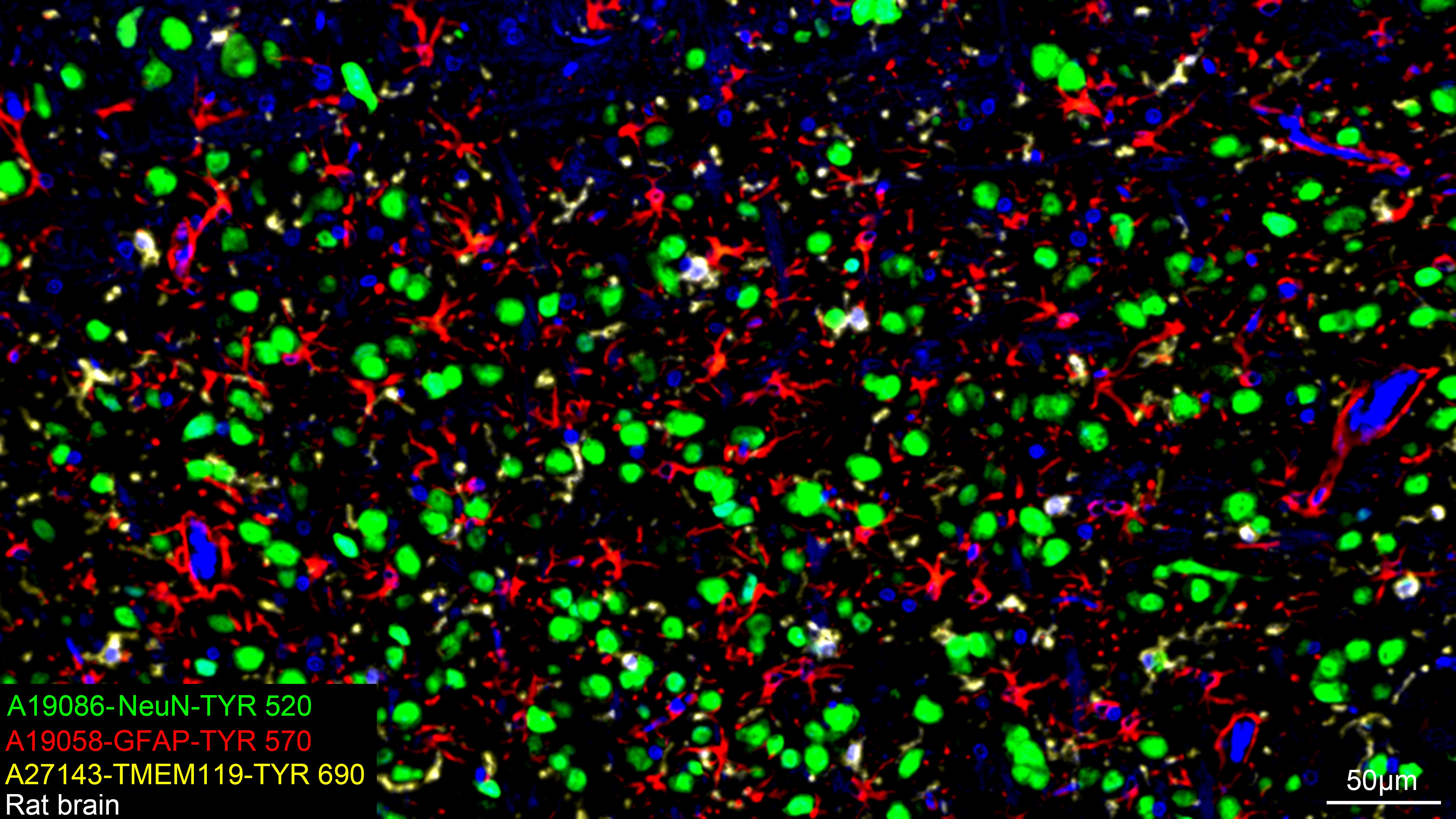
The multiplex IHC analysis on paraffin-embedded Rat brain tissue using the following specific primary antibodies and tyramide signal amplification (TSA) reagents (RK05903) : NeuN Rabbit mAb (CAB19086, 1:2000) with TSA-TYR-520 (Green), GFAP Rabbit mAb (CAB19058, 1:500) with TSA-TYR-570 (Red), and TMEM119 Rabbit mAb (CAB27143, 1:600) with TSA-TYR-690 (Yellow). DAPI (Blue) was used for nuclear staining. Prior to multiplex IHC staining, high-pressure antigen retrieval was performed using 0.01M citrate buffer at pH 6.0. The analysis was completed using a 20x objective lens.