The HSP90B1 Antibody (CAB6272) is a high-quality antibody developed for reliable detection and analysis of target proteins. This antibody, produced in rabbits, exhibits high reactivity with human samples and is validated for use in Western blot applications.HSP90B1 plays a crucial role in maintaining protein homeostasis and cell survival, making it a critical player in cellular stress response pathways. By targeting HSP90B1 with this polyclonal antibody, researchers can study its expression and activity in various cell types, shedding light on its function in normal cellular processes and disease states.
This antibody is validated for use in WB, IHC-P, IF/ICC, ELISA applications and has demonstrated reactivity against Human, Mouse, Rat samples.
Product Name:
HSP90B1 Antibody
SKU:
CAB6272
Size:
20μL, 100μL
Reactivity:
Human, Mouse, Rat
Conjugate:
Unconjugated
Immunogen:
Recombinant protein (or fragment).This information is considered to be commercially sensitive.
Recommended starting concentration is 1 μg/mL. Please optimize the concentration based on your specific assay requirements.
Synonyms:
ECGP, GP96, TRA1, GRP94, HEL35, HEL-S-125m, Grp94
Positive Sample:
MCF7, NIH/3T3, PC-12, Rat testis, PC-12, Rat testis, MCF7, NIH/3T3
Cellular Localization:
Endoplasmic Reticulum Lumen, Melanosome.
Calculated MW:
92kDa
Observed MW:
100kDa
This gene encodes a member of a family of adenosine triphosphate(ATP)-metabolizing molecular chaperones with roles in stabilizing and folding other proteins. The encoded protein is localized to melanosomes and the endoplasmic reticulum. Expression of this protein is associated with a variety of pathogenic states, including tumor formation. There is a microRNA gene located within the 5' exon of this gene. There are pseudogenes for this gene on chromosomes 1 and 15.
Purification Method
Affinity purification
Gene ID
7184
RRID
AB_2766877
Buffer Information
Store at -20℃. Avoid freeze / thaw cycles. Buffer: PBS containing 50% glycerol, preserved with proclin300 or sodium azide, pH 7.3.
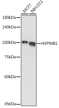
Western blot analysis of various lysates using Grp94 Rabbit pAb (CAB6272) at 1:1000 dilution. Secondary antibody: HRP-conjugated Goat anti-Rabbit IgG (H+L) (CABS014) at 1:10000 dilution. Lysates/proteins: 25μg per lane. Blocking buffer: 3% nonfat dry milk in TBST. Detection: ECL Basic Kit (AbGn00020). Exposure time: 30s.
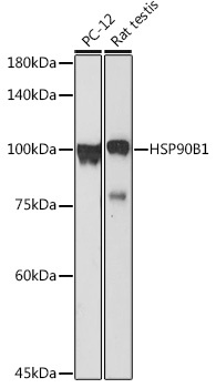
Western blot analysis of various lysates using Grp94 Rabbit pAb (CAB6272) at 1:1000 dilution. Secondary antibody: HRP-conjugated Goat anti-Rabbit IgG (H+L) (CABS014) at 1:10000 dilution. Lysates/proteins: 25μg per lane. Blocking buffer: 3% nonfat dry milk in TBST. Detection: ECL Basic Kit (AbGn00020). Exposure time: 180s.
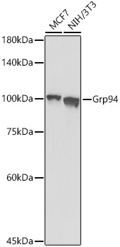
Western blot analysis of various lysates using Grp94 Rabbit pAb (CAB6272) at 1:1000 dilution incubated overnight at 4℃. Secondary antibody: HRP-conjugated Goat anti-Rabbit IgG (H+L) (CABS014) at 1:10000 dilution. Lysates/proteins: 25 μg per lane. Blocking buffer: 3% nonfat dry milk in TBST. Detection: ECL Basic Kit (AbGn00020). Exposure time: 30s.
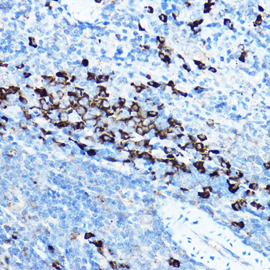
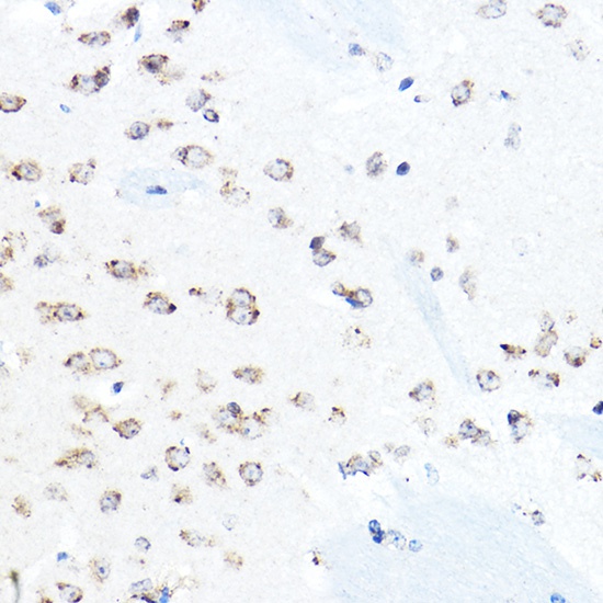
Immunohistochemistry analysis of paraffin-embedded Rat spleen using Grp94 Rabbit pAb (CAB6272) at dilution of 1:50 (40x lens). High pressure antigen retrieval performed with 0.01M Citrate buffer (pH 6.0) prior to IHC staining.
Immunohistochemistry analysis of paraffin-embedded Mouse brain using Grp94 Rabbit pAb (CAB6272) at dilution of 1:50 (40x lens). High pressure antigen retrieval performed with 0.01M Citrate buffer (pH 6.0) prior to IHC staining.
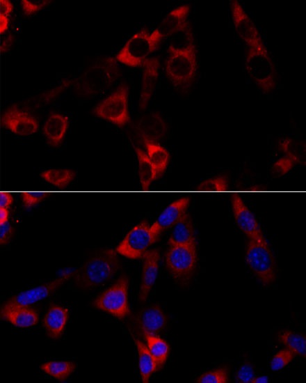
Immunofluorescence analysis of NIH-3T3 cells using Grp94 Rabbit pAb (CAB6272) at dilution of 1:100 (40x lens). Secondary antibody: Cy3-conjugated Goat anti-Rabbit IgG (H+L) (CABS007) at 1:500 dilution. Blue: DAPI for nuclear staining.