The IGF2BP2/IMP2 Antibody (CAB1774) is a high-quality antibody developed for reliable detection and analysis of target proteins. This antibody, raised in rabbits, exhibits high reactivity with human samples and is validated for use in various applications, including Western blotting.IGF2BP2, also known as insulin-like growth factor 2 mRNA-binding protein 2, plays a crucial role in post-transcriptional regulation of gene expression, particularly in the context of cell proliferation and differentiation.
This antibody is validated for use in WB, IF/ICC, ELISA, IF-P applications and has demonstrated reactivity against Human, Mouse, Rat samples.
Product Name:
IGF2BP2/IMP2 Antibody
SKU:
CAB1774
Size:
20μL, 100μL
Reactivity:
Human, Mouse, Rat
Conjugate:
Unconjugated
Immunogen:
Recombinant protein (or fragment).This information is considered to be commercially sensitive.
Recommended starting concentration is 1 μg/mL. Please optimize the concentration based on your specific assay requirements.
Synonyms:
IMP2, IMP-2, VICKZ2, IGF2BP2/IMP2
Positive Sample:
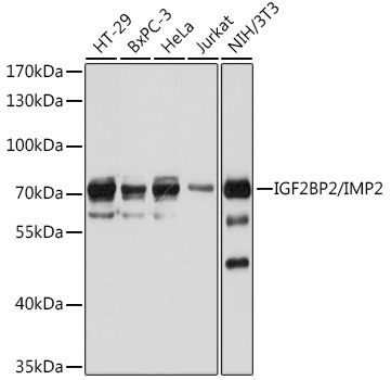
HT-29, BxPC-3, HeLa, Jurkat, NIH/3T3
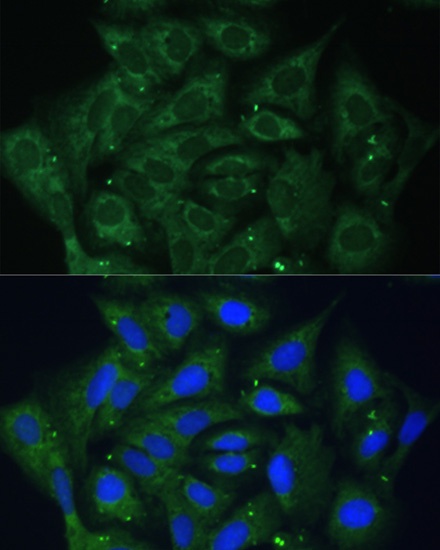
Cellular Localization:
Cytoplasm, Nucleus.
Calculated MW:
66kDa
Observed MW:
70kDa
This gene encodes a protein that binds the 5' UTR of insulin-like growth factor 2 (IGF2) mRNA and regulates its translation. It plays an important role in metabolism and variation in this gene is associated with susceptibility to diabetes. Alternative splicing and promoter usage results in multiple transcript variants. Related pseudogenes are found on several chromosomes.
Purification Method
Affinity purification
Gene ID
10644
RRID
AB_2763816
Buffer Information
Store at -20℃. Avoid freeze / thaw cycles. Buffer: PBS with 0.01% thimerosal,50% glycerol,pH7.3.
Western blot analysis of various lysates using IGF2BP2/IMP2 Rabbit pAb (CAB1774) at 1:1000 dilution. Secondary antibody: HRP-conjugated Goat anti-Rabbit IgG (H+L) (CABS014) at 1:10000 dilution. Lysates/proteins: 25μg per lane. Blocking buffer: 3% nonfat dry milk in TBST. Detection: ECL Basic Kit (AbGn00020). Exposure time: 30s.
Immunofluorescence analysis of U-2 OS cells using IGF2BP2/IMP2 Rabbit pAb (CAB1774) at dilution of 1:100. Secondary antibody: Cy3-conjugated Goat anti-Rabbit IgG (H+L) (CABS007) at 1:500 dilution. Blue: DAPI for nuclear staining.