The KAP1/TRIM28 Monoclonal Antibody (CAB19568) is a high-quality antibody developed for reliable detection and analysis of target proteins. The protein encoded by this gene mediates transcriptional control by interaction with the Kruppel-associated box repression domain found in many transcription factors. The protein localizes to the nucleus and is thought to associate with specific chromatin regions. The protein is a member of the tripartite motif family. This tripartite motif includes three zinc-binding domains, a RING, a B-box type 1 and a B-box type 2, and a coiled-coil region. RRID AB_2862673 Gene ID 10155 Swiss Prot Synonym KAP1; TF1B; RNF96; TIF1B; PPP1R157; TIF1beta; KAP1/TRIM28
This antibody is validated for use in WB, IHC-P, ELISA applications and has demonstrated reactivity against Human, Mouse, Rat samples.
Product Name:
KAP1/TRIM28 Monoclonal Antibody
SKU:
CAB19568
Size:
100μL, 20μL
Reactivity:
Human, Mouse, Rat
Clone Number:
ARC0047
Conjugate:
Unconjugated
Immunogen:
Synthetic peptide. This information is considered to be commercially sensitive.
Tested Applications:
WBIHC-PELISA
Recommended Dilution:
WB
1:1000 - 1:6000
IHC-P
1:200 - 1:2000
ELISA
Recommended starting concentration is 1 μg/mL. Please optimize the concentration based on your specific assay requirements.
The protein encoded by this gene mediates transcriptional control by interaction with the Kruppel-associated box repression domain found in many transcription factors. The protein localizes to the nucleus and is thought to associate with specific chromatin regions. The protein is a member of the tripartite motif family. This tripartite motif includes three zinc-binding domains, a RING, a B-box type 1 and a B-box type 2, and a coiled-coil region. RRID AB_2862673 Gene ID 10155 Swiss Prot Synonym KAP1; TF1B; RNF96; TIF1B; PPP1R157; TIF1beta; KAP1/TRIM28
Purification Method:
Affinity purification
Gene ID:
10155
RRID:
AB_2862673
Buffer Information:
Store at -20℃. Avoid freeze / thaw cycles. Buffer: PBS containing 50% glycerol and 0.05% BSA, preserved with proclin300 or sodium azide, pH 7.3.
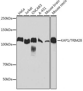
Western blot analysis of various lysates using KAP1/TRIM28 Rabbit mAb (CAB19568) at 1:1000 dilution. Secondary antibody: HRP-conjugated Goat anti-Rabbit IgG (H+L) (AS014) at 1:10000 dilution. Lysates/proteins: 25μg per lane. Blocking buffer: 3% nonfat dry milk in TBST. Detection: ECL Basic Kit (AbGn00020). Exposure time: 10s.
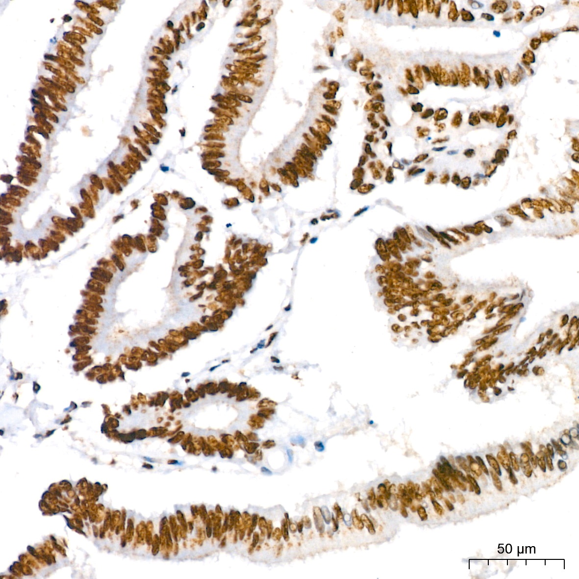
Immunohistochemistry analysis of paraffin-embedded Human colon carcinoma tissue using KAP1/TRIM28 Rabbit mAb (CAB19568) at dilution of 1:200 (40x lens). High pressure antigen retrieval performed with 0.01M Citrate Buffer (pH 6.0) prior to IHC staining.
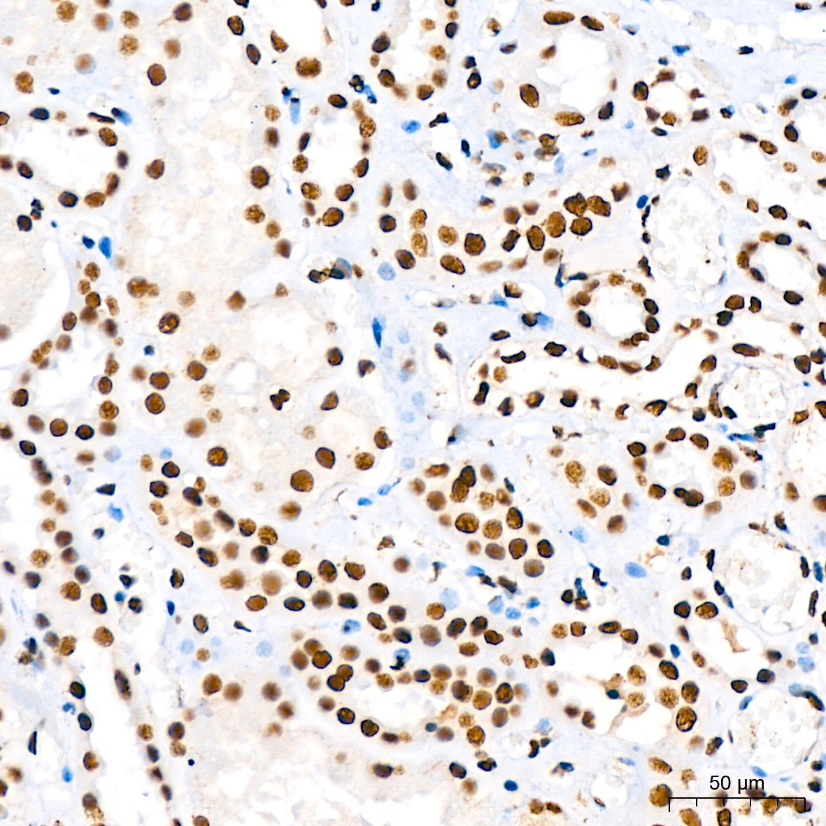
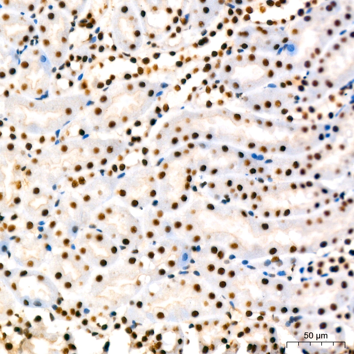
Immunohistochemistry analysis of paraffin-embedded Human kidney tissue using KAP1/TRIM28 Rabbit mAb (CAB19568) at dilution of 1:200 (40x lens). High pressure antigen retrieval performed with 0.01M Citrate Buffer (pH 6.0) prior to IHC staining.
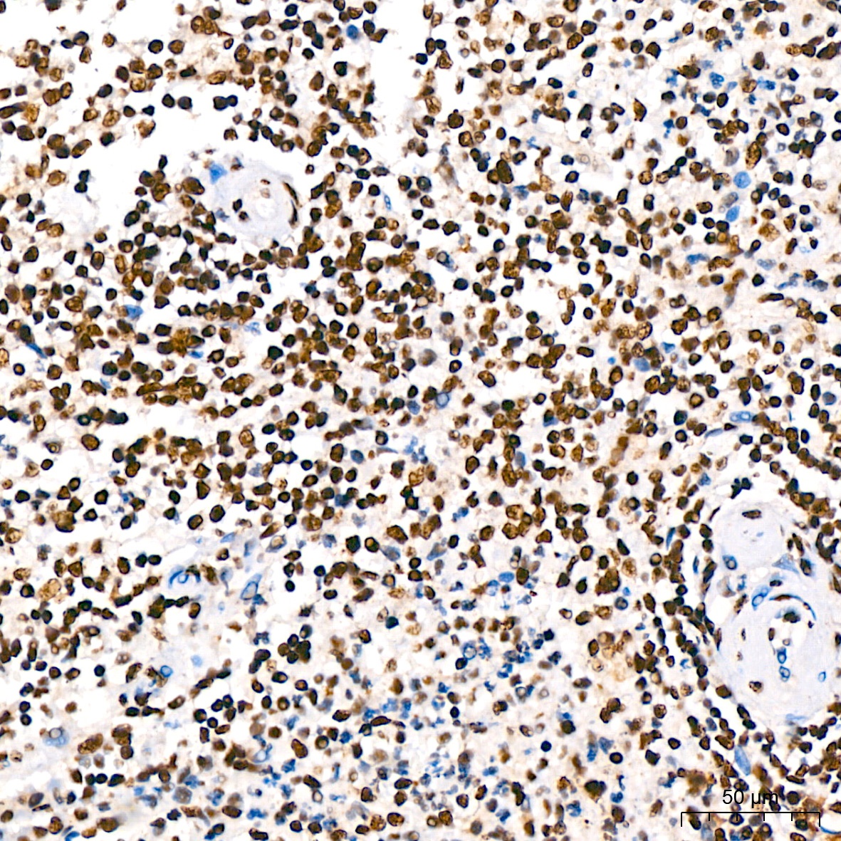
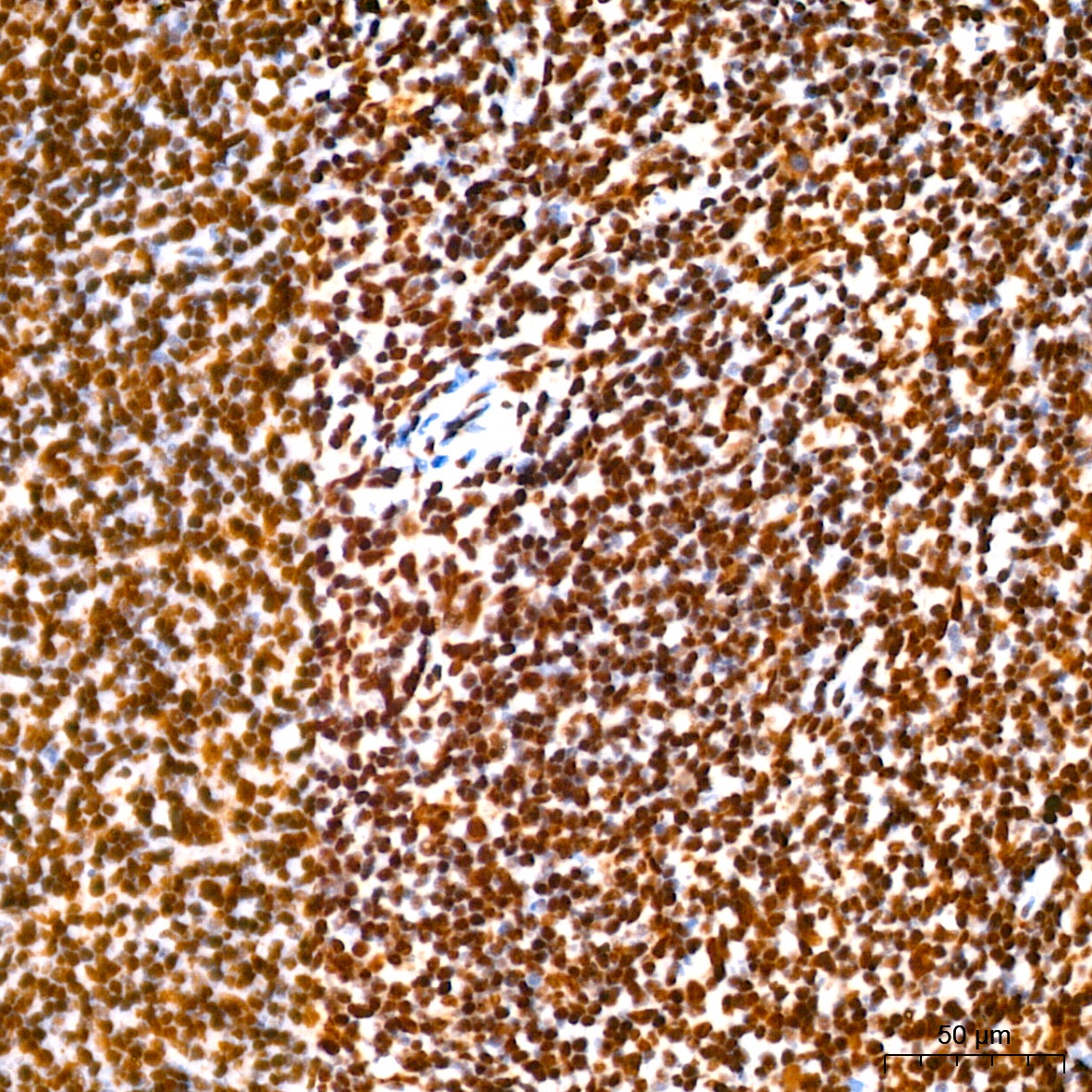
Immunohistochemistry analysis of paraffin-embedded Human spleen tissue using KAP1/TRIM28 Rabbit mAb (CAB19568) at dilution of 1:200 (40x lens). High pressure antigen retrieval performed with 0.01M Citrate Buffer (pH 6.0) prior to IHC staining.
Immunohistochemistry analysis of paraffin-embedded Mouse kidney tissue using KAP1/TRIM28 Rabbit mAb (CAB19568) at dilution of 1:200 (40x lens). High pressure antigen retrieval performed with 0.01M Citrate Buffer (pH 6.0) prior to IHC staining.
Immunohistochemistry analysis of paraffin-embedded Mouse spleen tissue using KAP1/TRIM28 Rabbit mAb (CAB19568) at dilution of 1:200 (40x lens). High pressure antigen retrieval performed with 0.01M Citrate Buffer (pH 6.0) prior to IHC staining.