The [KO Validated] Fascin/FSCN1 Antibody (CAB1904) is a high-quality antibody developed for reliable detection and analysis of target proteins. This antibody, produced in rabbits, has been rigorously tested and validated for use in various applications, particularly immunofluorescence and immunohistochemistry.Fascin is a critical regulator of cell migration and adhesion, playing a vital role in processes such as embryonic development, wound healing, and cancer metastasis. By targeting fascin with this antibody, researchers can investigate its expression and localization in different cell types and tissues, providing valuable insights into its functions and potential roles in disease progression.
This antibody is validated for use in WB, IHC-P, IF/ICC, ELISA applications and has demonstrated reactivity against Human, Mouse, Rat samples.
Product Name:
[KO Validated] Fascin/FSCN1 Antibody
SKU:
CAB1904
Size:
20μL, 100μL
Reactivity:
Human, Mouse, Rat
Conjugate:
Unconjugated
Immunogen:
Recombinant protein (or fragment).This information is considered to be commercially sensitive.
This gene encodes a member of the fascin family of actin-binding proteins. Fascin proteins organize F-actin into parallel bundles, and are required for the formation of actin-based cellular protrusions. The encoded protein plays a critical role in cell migration, motility, adhesion and cellular interactions. Expression of this gene is known to be regulated by several microRNAs, and overexpression of this gene may play a role in the metastasis of multiple types of cancer by increasing cell motility. Expression of this gene is also a marker for Reed-Sternberg cells in Hodgkin's lymphoma. A pseudogene of this gene is located on the long arm of chromosome 15.
Purification Method
Affinity purification
Gene ID
6624
RRID
AB_2862532
Buffer Information
Store at -20℃. Avoid freeze / thaw cycles. Buffer: PBS containing 50% glycerol, preserved with proclin300 or sodium azide, pH 7.3.
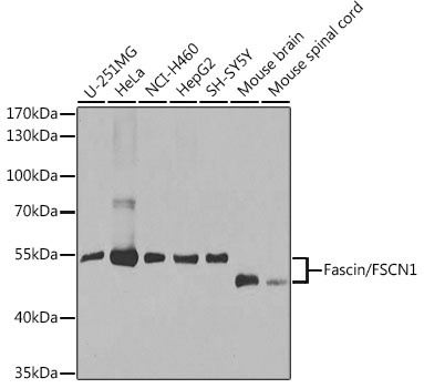
Western blot analysis of various lysates using [KO Validated] Fascin/FSCN1 Rabbit pAb (CAB1904) at 1:1000 dilution. Secondary antibody: HRP-conjugated Goat anti-Rabbit IgG (H+L) (CABS014) at 1:10000 dilution. Lysates/proteins: 25μg per lane. Blocking buffer: 3% nonfat dry milk in TBST. Detection: ECL Basic Kit (AbGn00020). Exposure time: 30s.
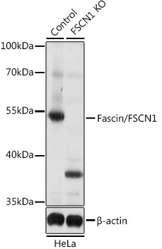
Western blot analysis of lysates from wild type (WT) and Fascin/Fascin/FSCN1 knockout (KO) HeLa cells, using [KO Validated] Fascin/FSCN1 Rabbit pAb (CAB1904) at 1:1000 dilution. Secondary antibody: HRP-conjugated Goat anti-Rabbit IgG (H+L) (CABS014) at 1:10000 dilution. Lysates/proteins: 25μg per lane. Blocking buffer: 3% nonfat dry milk in TBST. Detection: ECL Basic Kit (AbGn00020). Exposure time: 5s.
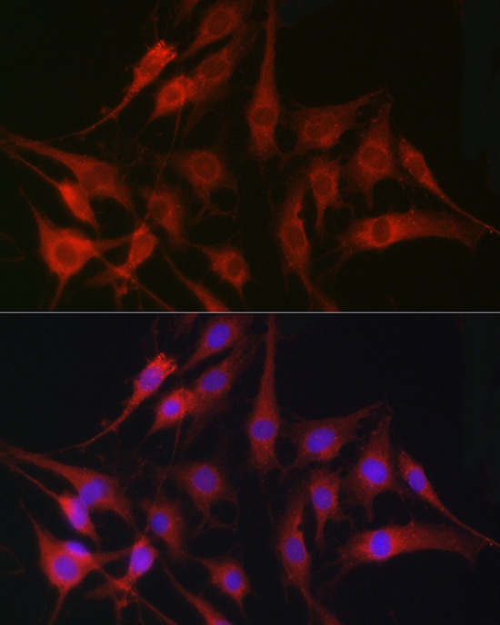
Immunofluorescence analysis of C6 cells using [KO Validated] Fascin/Fascin/FSCN1 Rabbit pAb (CAB1904) at dilution of 1:100 (40x lens). Secondary antibody: Cy3-conjugated Goat anti-Rabbit IgG (H+L) (CABS007) at 1:500 dilution. Blue: DAPI for nuclear staining.
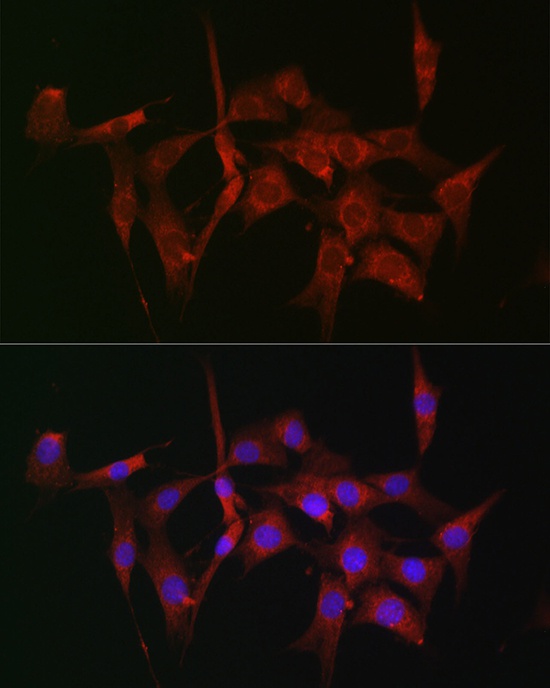
Immunofluorescence analysis of NIH-3T3 cells using [KO Validated] Fascin/Fascin/FSCN1 Rabbit pAb (CAB1904) at dilution of 1:100 (40x lens). Secondary antibody: Cy3-conjugated Goat anti-Rabbit IgG (H+L) (CABS007) at 1:500 dilution. Blue: DAPI for nuclear staining.