The [KO Validated] Integrin alpha 2 (ITGA2/CD49b) Antibody (CAB7629) is a high-quality antibody developed for reliable detection and analysis of target proteins. This antibody, developed using rabbits, exhibits high reactivity with human samples and has been validated for use in Western blot applications.Integrin alpha 2 plays a crucial role in cell-matrix interactions, controlling cell migration, proliferation, and differentiation. Its involvement in various biological processes such as wound healing, angiogenesis, and tumor progression makes it a key target for investigation in the fields of cancer research and vascular biology.
This antibody is validated for use in WB, IHC-P, IF/ICC, ELISA applications and has demonstrated reactivity against Human, Mouse, Rat samples.
Recommended starting concentration is 1 μg/mL. Please optimize the concentration based on your specific assay requirements.
Synonyms:
BR, GPIa, CD49B, HPA-5, VLA-2, VLAA2, b)
Positive Sample:
293T, HeLa, HT-1080
Cellular Localization:
Membrane, Single-Pass Type I Membrane Protein.
Calculated MW:
129kDa
Observed MW:
150kDa
This gene encodes the alpha subunit of a transmembrane receptor for collagens and related proteins. The encoded protein forms a heterodimer with a beta subunit and mediates the adhesion of platelets and other cell types to the extracellular matrix. Loss of the encoded protein is associated with bleeding disorder platelet-type 9. Antibodies against this protein are found in several immune disorders, including neonatal alloimmune thrombocytopenia. This gene is located adjacent to a related alpha subunit gene. Alternative splicing results in multiple transcript variants.
Purification Method
Affinity purification
Gene ID
3673
RRID
AB_2863553
Buffer Information
Store at -20℃. Avoid freeze / thaw cycles. Buffer: PBS containing 50% glycerol, preserved with proclin300 or sodium azide, pH 7.3.
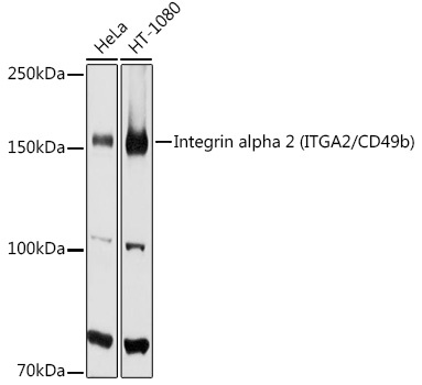
Western blot analysis of various lysates using [KO Validated] Integrin alpha 2 (ITGA2/CD49b) Rabbit pAb (CAB7629) at 1:1000 dilution. Secondary antibody: HRP-conjugated Goat anti-Rabbit IgG (H+L) (CABS014) at 1:10000 dilution. Lysates/proteins: 25μg per lane. Blocking buffer: 3% nonfat dry milk in TBST. Detection: ECL Basic Kit (AbGn00020). Exposure time: 90s.
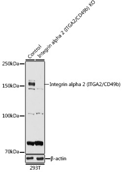
Western blot analysis of lysates from wild type (WT) and Integrin alpha 2 (ITGA2/CD49b) knockout (KO) 293T cells, using [KO Validated] Integrin alpha 2 (ITGA2/CD49b) Rabbit pAb (CAB7629) at 1:1000 dilution. Secondary antibody: HRP-conjugated Goat anti-Rabbit IgG (H+L) (CABS014) at 1:10000 dilution. Lysates/proteins: 25μg per lane. Blocking buffer: 3% nonfat dry milk in TBST. Detection: ECL Basic Kit (AbGn00020). Exposure time: 90s.
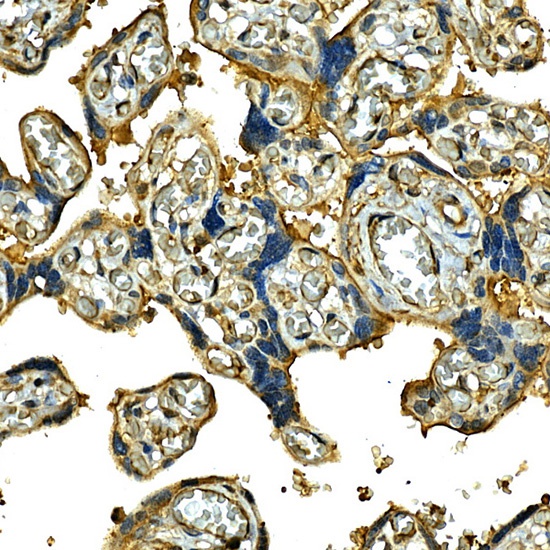
Immunohistochemistry analysis of paraffin-embedded Human placenta using [KO Validated] Integrin alpha 2 (ITGA2/CD49b) Rabbit pAb (CAB7629) at dilution of 1:50 (40x lens). High pressure antigen retrieval performed with 0.01M Citrate buffer (pH 6.0) prior to IHC staining.
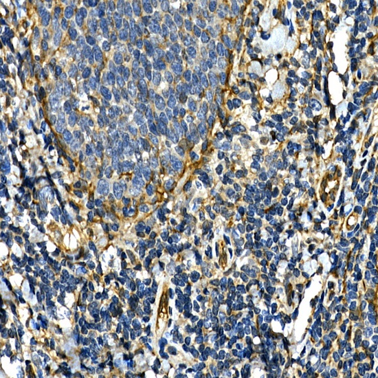
Immunohistochemistry analysis of paraffin-embedded Human tonsil using [KO Validated] Integrin alpha 2 (ITGA2/CD49b) Rabbit pAb (CAB7629) at dilution of 1:50 (40x lens). High pressure antigen retrieval performed with 0.01M Citrate buffer (pH 6.0) prior to IHC staining.