The [KO Validated] TTC11/FIS1 Monoclonal Antibody (CAB19666) is a high-quality antibody developed for reliable detection and analysis of target proteins. Enables identical protein binding activity. Involved in several processes, including calcium-mediated signaling using intracellular calcium source; cellular calcium ion homeostasis; and mitochondrion organization. Acts upstream of or within mitochondrion morphogenesis. Located in mitochondrion and peroxisome. Is integral component of mitochondrial outer membrane and integral component of peroxisomal membrane. Part of protein-containing complex. Biomarker of Alzheimer's disease.
This antibody is validated for use in WB, IHC-P, IF/ICC, IP, ELISA applications and has demonstrated reactivity against Human, Mouse, Rat samples.
Product Name:
[KO Validated] TTC11/FIS1 Monoclonal Antibody
SKU:
CAB19666
Size:
100μL, 20μL
Reactivity:
Human, Mouse, Rat
Clone Number:
ARC5010-03
Conjugate:
Unconjugated
Immunogen:
Recombinant protein (or fragment).This information is considered to be commercially sensitive.
Tested Applications:
WBIHC-PIF/ICCIPELISA
Recommended Dilution:
WB
1:1000 - 1:6000
IHC-P
1:100 - 1:400
IF/ICC
1:100 - 1:400
IP
0.5μg-4μg antibody for 200μg-400μg extracts of whole cells
ELISA
Recommended starting concentration is 1 μg/mL. Please optimize the concentration based on your specific assay requirements.
Enables identical protein binding activity. Involved in several processes, including calcium-mediated signaling using intracellular calcium source; cellular calcium ion homeostasis; and mitochondrion organization. Acts upstream of or within mitochondrion morphogenesis. Located in mitochondrion and peroxisome. Is integral component of mitochondrial outer membrane and integral component of peroxisomal membrane. Part of protein-containing complex. Biomarker of Alzheimer's disease.
Purification Method
Affinity purification
Gene ID
51024
RRID
AB_2862724
Buffer Information
Store at -20℃. Avoid freeze / thaw cycles. Buffer: PBS containing 50% glycerol and 0.05% BSA, preserved with proclin300 or sodium azide, pH 7.3.
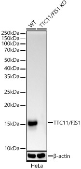
Western blot analysis of lysates from wild type (WT) and TTC11/FIS1 knockout (KO) HeLa(KO) cells, using [KO Validated] TTC11/FIS1 Rabbit mAb (CAB19666) at 1:1000 dilution incubated overnight at 4℃. Secondary antibody: HRP-conjugated Goat anti-Rabbit IgG (H+L) (AS014) at 1:10000 dilution. Lysates/proteins: 25μg per lane. Blocking buffer: 3% nonfat dry milk in TBST. Detection: ECL Basic Kit (AbGn00020). Exposure time: 10s.
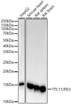
Western blot analysis of various lysates, using [KO Validated] TTC11/FIS1 Rabbit mAb (CAB19666) at 1:1000 dilution incubated overnight at 4℃. Secondary antibody: HRP-conjugated Goat anti-Rabbit IgG (H+L) (AS014) at 1:10000 dilution. Lysates/proteins: 25μg per lane. Blocking buffer: 3% nonfat dry milk in TBST. Detection: ECL Basic Kit (AbGn00020). Exposure time: 10s.
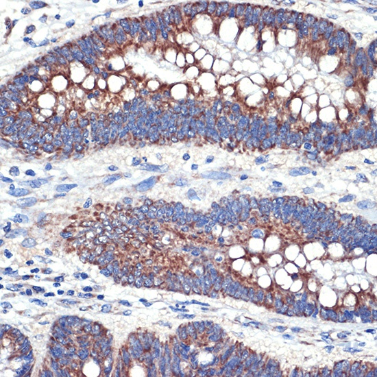
Immunohistochemistry analysis of paraffin-embedded Human colon carcinoma tissue using [KO Validated] TTC11/FIS1 Rabbit mAb (CAB19666) at a dilution of 1:100 (40x lens). Microwave antigen retrieval performed with 0.01M Tris-EDTA Buffer (pH 9.0) prior to IHC staining.
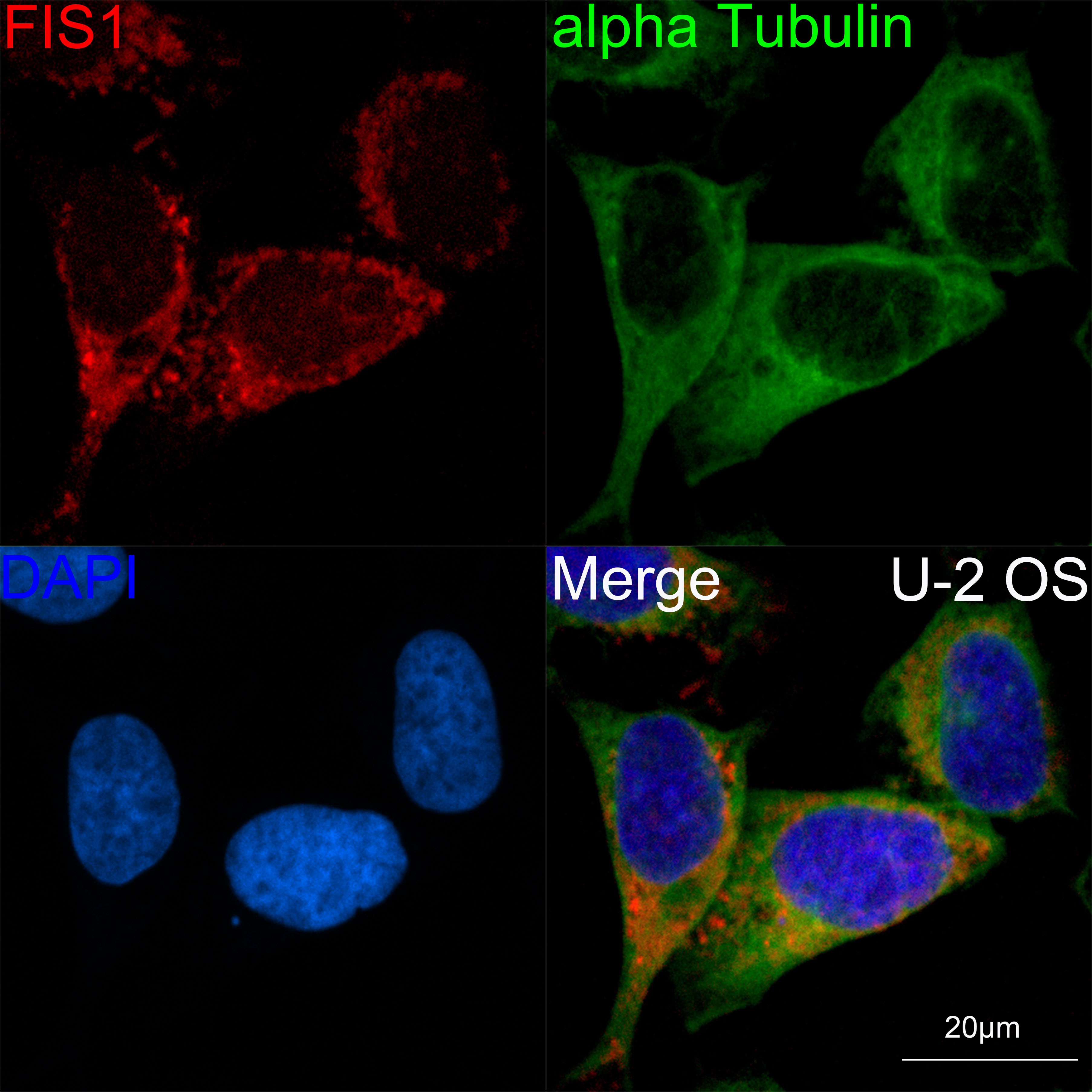
Confocal imaging of U-2 OS cells using [KO Validated] TTC11/FIS1 Rabbit mAb (CAB19666,dilution 1:100)(Red). The cells were counterstained with α-Tubulin Mouse mAb (AC012,dilution 1:400) (Green). DAPI was used for nuclear staining (blue). Objective: 60x.