The [KO Validated] Vimentin Monoclonal Antibody (CAB19607) is a high-quality antibody developed for reliable detection and analysis of target proteins. This gene encodes a type III intermediate filament protein. Intermediate filaments, along with microtubules and actin microfilaments, make up the cytoskeleton. The encoded protein is responsible for maintaining cell shape and integrity of the cytoplasm, and stabilizing cytoskeletal interactions. This protein is involved in neuritogenesis and cholesterol transport and functions as an organizer of a number of other critical proteins involved in cell attachment, migration, and signaling. Bacterial and viral pathogens have been shown to attach to this protein on the host cell surface. Mutations in this gene are associated with congenital cataracts in human patients. RRID AB_2862696 Gene ID 7431 Swiss Prot Synonym CTRCT30; HEL113; Vimentin; VIM; vimentin; in
This antibody is validated for use in WB, IHC-P, IF/ICC, IP, ELISA, IF-P applications and has demonstrated reactivity against Human, Mouse, Rat samples.
Product Name:
[KO Validated] Vimentin Monoclonal Antibody
SKU:
CAB19607
Size:
100μL, 20μL
Reactivity:
Human, Mouse, Rat
Clone Number:
ARC0086
Conjugate:
Unconjugated
Immunogen:
Synthetic peptide. This information is considered to be commercially sensitive.
Tested Applications:
WBIHC-PIF/ICCIPELISAIF-P
Recommended Dilution:
WB
1:5000 - 1:40000
IP
0.5μg-4μg antibody for 200μg-400μg extracts of whole cells
IF
/
ICC
1:200 - 1:2000
IF-P
1:200 - 1:2000
IHC-P
1:1000 - 1:4000
ELISA
Recommended starting concentration is 1 μg/mL. Please optimize the concentration based on your specific assay requirements.
Synonyms:
CTRCT30, HEL113, Vimentin, VIM, vimentin, in
Positive Sample:
293T, HeLa
Cellular Localization:
Cytoplasm.
Calculated MW:
54kDa
Observed MW:
60kDa
This gene encodes a type III intermediate filament protein. Intermediate filaments, along with microtubules and actin microfilaments, make up the cytoskeleton. The encoded protein is responsible for maintaining cell shape and integrity of the cytoplasm, and stabilizing cytoskeletal interactions. This protein is involved in neuritogenesis and cholesterol transport and functions as an organizer of a number of other critical proteins involved in cell attachment, migration, and signaling. Bacterial and viral pathogens have been shown to attach to this protein on the host cell surface. Mutations in this gene are associated with congenital cataracts in human patients. RRID AB_2862696 Gene ID 7431 Swiss Prot Synonym CTRCT30; HEL113; Vimentin; VIM; vimentin; in
Purification Method:
Affinity purification
Gene ID:
7431
RRID:
AB_2862696
Buffer Information:
Store at -20℃. Avoid freeze / thaw cycles. Buffer: PBS containing 50% glycerol and 0.05% BSA, preserved with proclin300 or sodium azide, pH 7.3.
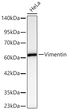
Western blot analysis of lysates from HeLa cells using [KD Validated] Vimentin Rabbit mAb (CAB19607) at 1:5000 dilution incubated overnight at 4℃. Secondary antibody: HRP-conjugated Goat anti-Rabbit IgG (H+L) (AS014) at 1:10000 dilution. Lysates/proteins: 25 μg per lane. Blocking buffer: 3% nonfat dry milk in TBST. Detection: ECL Basic Kit (AbGn00020). Exposure time: 0.5s.
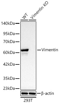
Western blot analysis of lysates from wild type (WT) and Vimentin knockout (KO) 293T cells using [KD Validated] Vimentin Rabbit mAb (CAB19607) at 1:5500 dilution incubated overnight at 4℃. Secondary antibody: HRP-conjugated Goat anti-Rabbit IgG (H+L) (AS014) at 1:10000 dilution. Lysates/proteins: 25 μg per lane. Blocking buffer: 3% nonfat dry milk in TBST. Detection: ECL Basic Kit (AbGn00020). Exposure time: 0.5s.
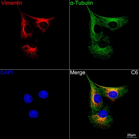
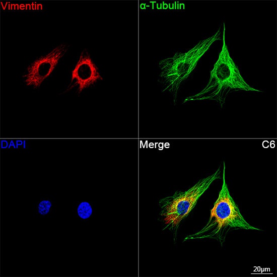
Confocal imaging of C6 cells using [KD Validated] Vimentin Rabbit mAb (CAB19607,dilution 1:200) followed by a further incubation with Cy3 Goat Anti-Rabbit IgG (H+L) (AS007, dilution 1:500) (Red). The cells were counterstained with α-Tubulin Mouse mAb (AC012, dilution 1:400) followed by incubation with ABflo® 488-conjugated Goat Anti-Mouse IgG (H+L) Ab (AS076, dilution 1:500) (Green). DAPI was used for nuclear staining (Blue). Objective: 100x.
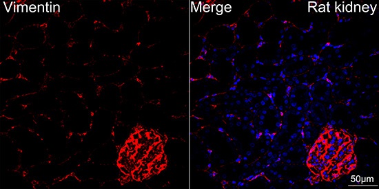
Confocal imaging of paraffin-embedded Rat kidney tissue using [KD Validated] Vimentin Rabbit mAb (CAB19607, dilution 1:200) followed by a further incubation with Cy3 Goat Anti-Rabbit IgG (H+L) (AS007, dilution 1:500) (Red). DAPI was used for nuclear staining (Blue). High pressure antigen retrieval performed with 0.01M Citrate Buffer (pH 6.0) prior to IF staining. Objective: 40x.
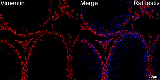
Confocal imaging of paraffin-embedded Rat testis tissue using [KD Validated] Vimentin Rabbit mAb (CAB19607, dilution 1:200) followed by a further incubation with Cy3 Goat Anti-Rabbit IgG (H+L) (AS007, dilution 1:500) (Red). DAPI was used for nuclear staining (Blue). High pressure antigen retrieval performed with 0.01M Citrate Buffer (pH 6.0) prior to IF staining. Objective: 40x.
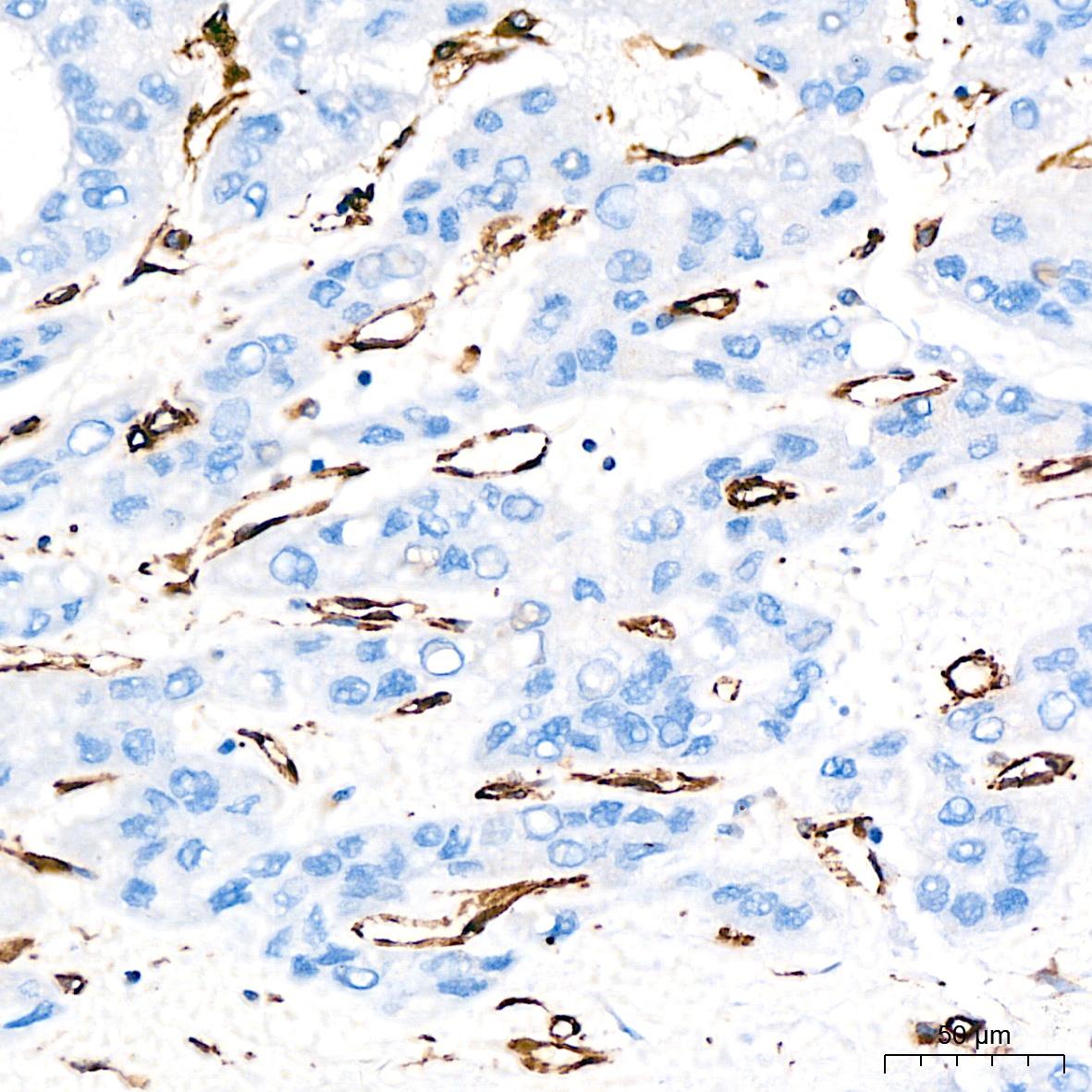
Immunohistochemistry analysis of paraffin-embedded Human colon carcinoma tissue using [KD Validated] Vimentin Rabbit mAb (CAB19607) at a dilution of 1:1600 (40x lens). High pressure antigen retrieval performed with 0.01M Citrate Buffer(pH 6.0) prior to IHC staining.
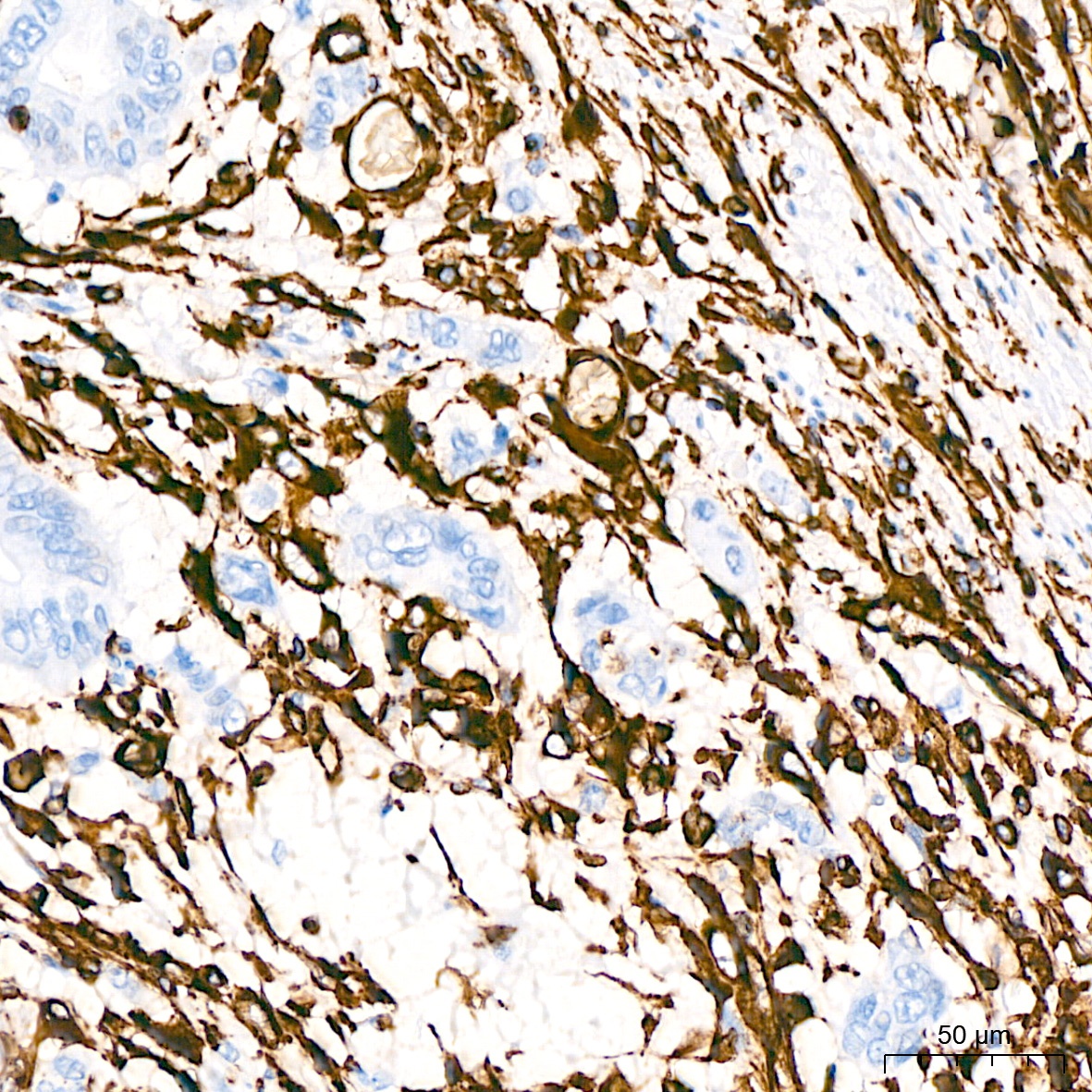
Immunohistochemistry analysis of paraffin-embedded Human liver cancer tissue using [KD Validated] Vimentin Rabbit mAb (CAB19607) at a dilution of 1:1600 (40x lens). High pressure antigen retrieval performed with 0.01M Citrate Buffer(pH 6.0) prior to IHC staining.
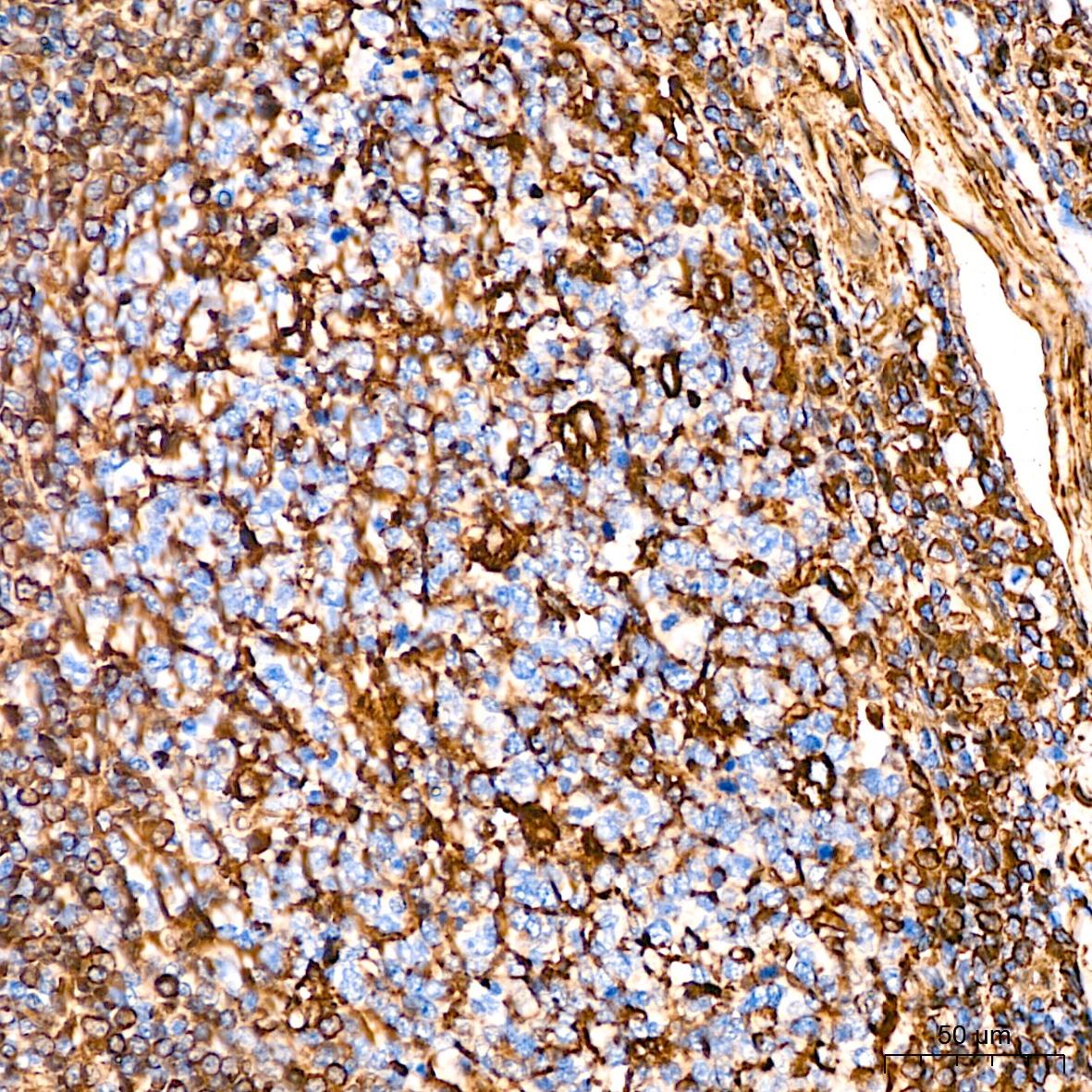
Immunohistochemistry analysis of paraffin-embedded Human tonsil tissue using [KD Validated] Vimentin Rabbit mAb (CAB19607) at a dilution of 1:1600 (40x lens). High pressure antigen retrieval performed with 0.01M Citrate Buffer(pH 6.0) prior to IHC staining.
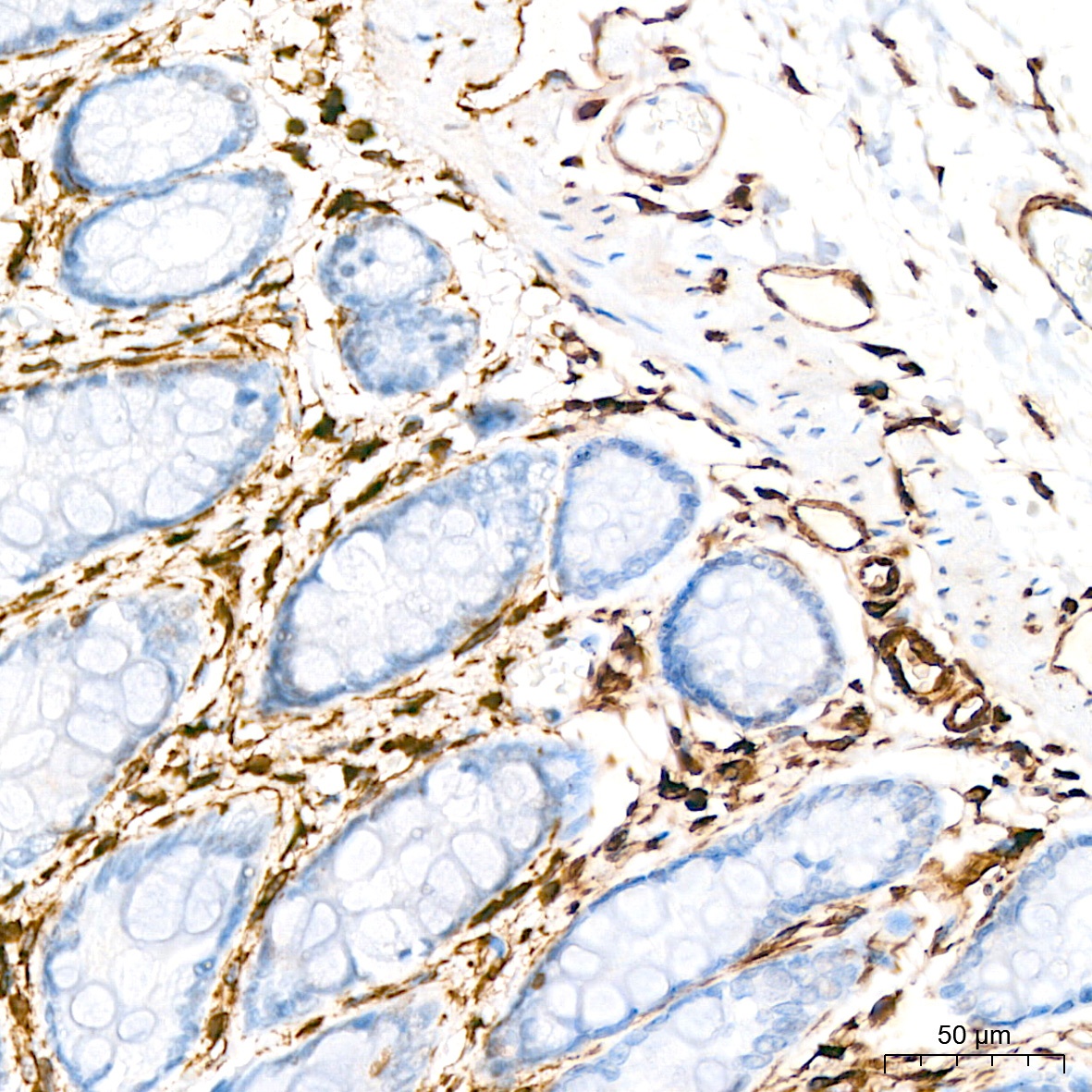
Immunohistochemistry analysis of paraffin-embedded Mouse colon tissue using [KD Validated] Vimentin Rabbit mAb (CAB19607) at a dilution of 1:1600 (40x lens). High pressure antigen retrieval performed with 0.01M Citrate Buffer(pH 6.0) prior to IHC staining.
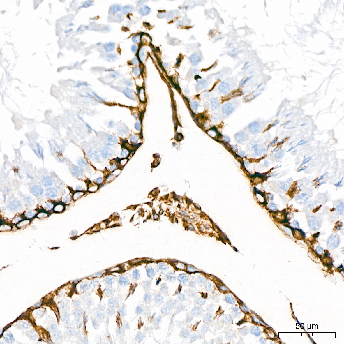
Immunohistochemistry analysis of paraffin-embedded Rat testis tissue using [KD Validated] Vimentin Rabbit mAb (CAB19607) at a dilution of 1:1600 (40x lens). High pressure antigen retrieval performed with 0.01M Citrate Buffer(pH 6.0) prior to IHC staining.
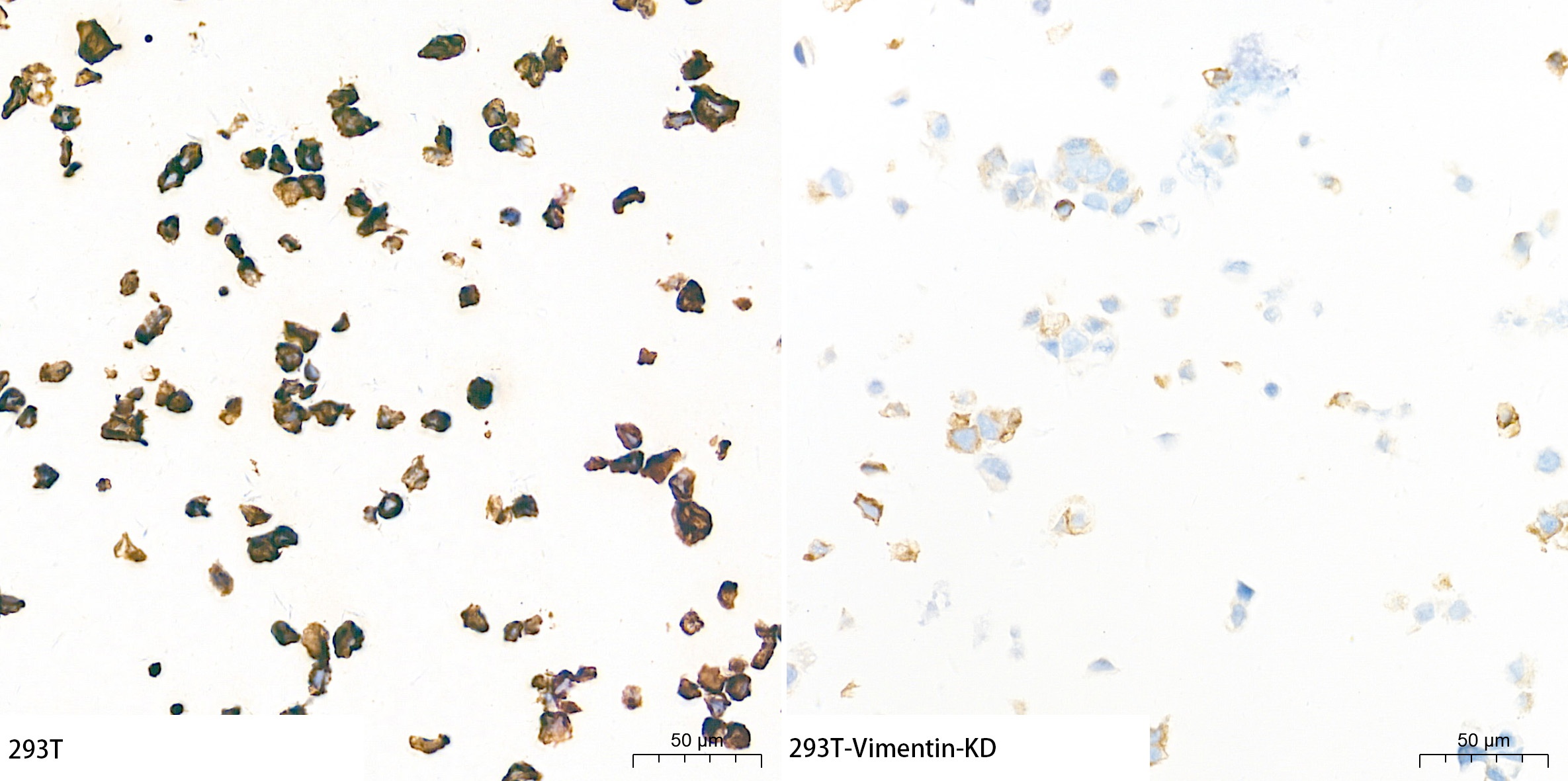
Immunohistochemistry analysis of paraffin-embedded 293T and 293T-VIM-KD cells using [KD Validated] Vimentin Rabbit mAb (CAB19607) at a dilution of 1:1600 (40x lens). High pressure antigen retrieval performed with 0.01M Tris-EDTA Buffer (pH 9.0) prior to IHC staining.
Confocal imaging of C6 cells using [KD Validated] Vimentin Rabbit mAb (CAB19607, dilution 1:700) followed by a further incubation with Cy3 Goat Anti-Rabbit IgG (H+L) (AS007, dilution 1:500) (Red). The cells were counterstained with α-Tubulin Mouse mAb (AC012, dilution 1:400) followed by incubation with ABflo® 488-conjugated Goat Anti-Mouse IgG (H+L) Ab (AS076, dilution 1:500) (Green). DAPI was used for nuclear staining (Blue). Objective: 100x.
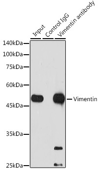
Immunoprecipitation analysis of 300 μg extracts of Jurkat cells using 3 μg [KD Validated] Vimentin Rabbit mAb (CAB19607). Western blot was performed from the immunoprecipitate using [KD Validated] Vimentin Rabbit mAb (CAB19607) at a dilution of 1:1000.