The Matrin 3 Monoclonal Antibody (CAB1027) is a high-quality antibody developed for reliable detection and analysis of target proteins. This polyclonal antibody, derived from rabbits, displays high reactivity with human samples and is suitable for use in Western blot applications. By specifically binding to MATRIN-3, this antibody allows for the detection and analysis of this protein in various cellular contexts, making it an essential asset for investigations in molecular biology and disease pathology.MATRIN-3 is known for its role in RNA splicing and transport, as well as its involvement in nuclear matrix organization. Dysregulation of MATRIN-3 has been linked to neurological disorders, including amyotrophic lateral sclerosis (ALS) and frontotemporal dementia (FTD).
This antibody is validated for use in WB, IHC-P, IF/ICC, ELISA applications and has demonstrated reactivity against Human, Mouse, Rat samples.
Product Name:
Matrin 3 Monoclonal Antibody
SKU:
CAB1027
Size:
20μL, 100μL
Reactivity:
Human, Mouse, Rat
Clone Number:
ARC1854
Conjugate:
Unconjugated
Immunogen:
Synthetic peptide. This information is considered to be commercially sensitive.
Recommended starting concentration is 1 μg/mL. Please optimize the concentration based on your specific assay requirements.
Synonyms:
MPD2, ALS21, VCPDM, Matrin 3
Positive Sample:
293T, K-562, Mouse brain, Rat brain
Cellular Localization:
Nucleus Matrix.
Calculated MW:
95kDa
Observed MW:
125kDa
This gene encodes a nuclear matrix protein, which is proposed to stabilize certain messenger RNA species. Mutations of this gene are associated with distal myopathy 2, which often includes vocal cord and pharyngeal weakness. Alternatively spliced transcript variants, including read-through transcripts composed of the upstream small nucleolar RNA host gene 4 (non-protein coding) and matrin 3 gene sequence, have been identified. Pseudogenes of this gene are located on chromosomes 1 and X.
Purification Method
Affinity purification
Gene ID
9782
RRID
AB_2861490
Buffer Information
Store at -20℃. Avoid freeze / thaw cycles. Buffer: PBS containing 50% glycerol and 0.05% BSA, preserved with proclin300 or sodium azide, pH 7.3.
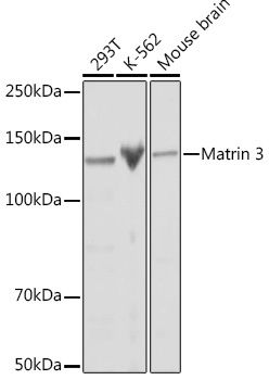
Western blot analysis of various lysates using Matrin 3 Rabbit mAb (CAB1027) at 1:1000 dilution. Secondary antibody: HRP-conjugated Goat anti-Rabbit IgG (H+L) (CABS014) at 1:10000 dilution. Lysates/proteins: 25μg per lane. Blocking buffer: 3% nonfat dry milk in TBST. Detection: ECL Basic Kit (AbGn00020). Exposure time: 10s.
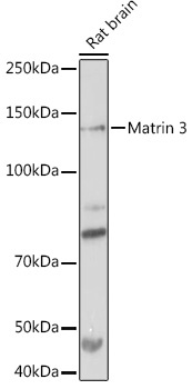
Western blot analysis of lysates from Rat brain, using Matrin 3 Rabbit mAb (CAB1027) at 1:1000 dilution. Secondary antibody: HRP-conjugated Goat anti-Rabbit IgG (H+L) (CABS014) at 1:10000 dilution. Lysates/proteins: 25μg per lane. Blocking buffer: 3% nonfat dry milk in TBST. Detection: ECL Basic Kit (AbGn00020). Exposure time: 30s.
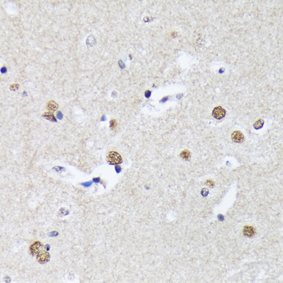
Immunohistochemistry analysis of paraffin-embedded Human brain using Matrin 3 Rabbit mAb (CAB1027) at dilution of 1:100 (40x lens). Microwave antigen retrieval performed with 0.01M Tris/EDTA Buffer (pH 9.0) prior to IHC staining.
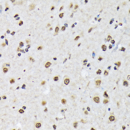
Immunohistochemistry analysis of paraffin-embedded Mouse spinal cord using Matrin 3 Rabbit mAb (CAB1027) at dilution of 1:100 (40x lens). Microwave antigen retrieval performed with 0.01M Tris/EDTA Buffer (pH 9.0) prior to IHC staining.
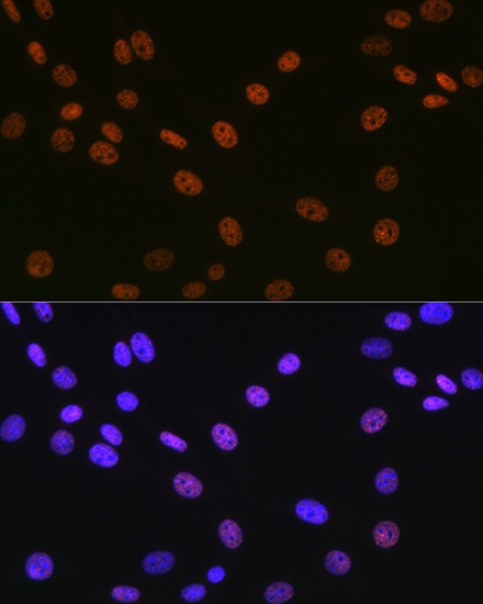
Immunofluorescence analysis of NIH-3T3 cells using Matrin 3 Rabbit mAb (CAB1027) at dilution of 1:100 (40x lens). Secondary antibody: Cy3-conjugated Goat anti-Rabbit IgG (H+L) (CABS007) at 1:500 dilution. Blue: DAPI for nuclear staining.
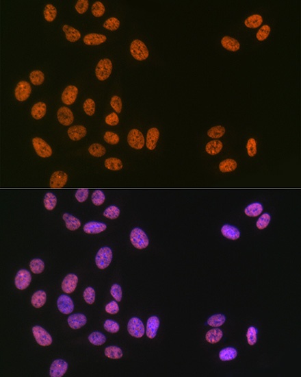
Immunofluorescence analysis of U-2 OS cells using Matrin 3 Rabbit mAb (CAB1027) at dilution of 1:100 (40x lens). Secondary antibody: Cy3-conjugated Goat anti-Rabbit IgG (H+L) (CABS007) at 1:500 dilution. Blue: DAPI for nuclear staining.