The MDC1 Antibody (CAB8358) is a high-quality antibody developed for reliable detection and analysis of target proteins. The protein encoded by this gene contains an N-terminal forkhead domain, two BRCA1 C-terminal (BRCT) motifs and a central domain with 13 repetitions of an approximately 41-amino acid sequence. The encoded protein is required to activate the intra-S phase and G2/M phase cell cycle checkpoints in response to DNA damage. This nuclear protein interacts with phosphorylated histone H2AX near sites of DNA double-strand breaks through its BRCT motifs, and facilitates recruitment of the ATM kinase and meiotic recombination 11 protein complex to DNA damage foci.
This antibody is validated for use in WB, IHC-P, IF/ICC, ELISA applications and has demonstrated reactivity against Human samples.
Product Name:
MDC1 Antibody
SKU:
CAB8358
Size:
100μL, 20μL
Reactivity:
Human
Conjugate:
Unconjugated
Immunogen:
Recombinant protein (or fragment).This information is considered to be commercially sensitive.
Tested Applications:
WBIHC-PIF/ICCELISA
Recommended Dilution:
WB
1:500 - 1:2000
IHC-P
1:50 - 1:200
IF/ICC
1:50 - 1:200
ELISA
Recommended starting concentration is 1 μg/mL. Please optimize the concentration based on your specific assay requirements.
Synonyms:
NFBD1, MDC1
Positive Sample:
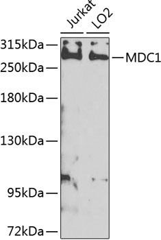
Jurkat, LO2
Cellular Localization:
Chromosome, Nucleus.
Calculated MW:
227kDa
Observed MW:
270kDa
The protein encoded by this gene contains an N-terminal forkhead domain, two BRCA1 C-terminal (BRCT) motifs and a central domain with 13 repetitions of an approximately 41-amino acid sequence. The encoded protein is required to activate the intra-S phase and G2/M phase cell cycle checkpoints in response to DNA damage. This nuclear protein interacts with phosphorylated histone H2AX near sites of DNA double-strand breaks through its BRCT motifs, and facilitates recruitment of the ATM kinase and meiotic recombination 11 protein complex to DNA damage foci.
Purification Method
Affinity purification
Gene ID
9656
RRID
AB_2770323
Buffer Information
Store at -20℃. Avoid freeze / thaw cycles. Buffer: PBS containing 50% glycerol, preserved with proclin300 or sodium azide, pH 7.3.
Western blot analysis of various lysates using MDC1 Rabbit pAb (CAB8358) at 1:1000 dilution._Secondary antibody: HRP-conjugated Goat anti-Rabbit IgG (H+L) (AS014) at 1:10000 dilution._Lysates/proteins: 25μg per lane._Blocking buffer: 3% nonfat dry milk in TBST._Detection: ECL Enhanced Kit (AbGn00021)._Exposure time: 5s.
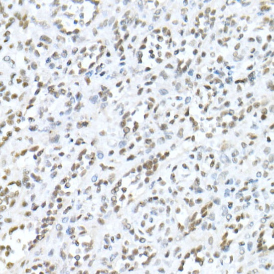
Immunohistochemistry analysis of paraffin-embedded Human spleen using MDC1 Rabbit pAb (CAB8358) at dilution of 1:20 (40x lens). High pressure antigen retrieval performed with 0.01M Citrate buffer (pH 6.0) prior to IHC staining.
Immunofluorescence analysis of U-2 OS cells using MDC1 Rabbit pAb (CAB8358) at dilution of 1:100. Secondary antibody: Cy3-conjugated Goat anti-Rabbit IgG (H+L) (AS007) at 1:500 dilution. Blue: DAPI for nuclear staining.