The MSRB1 Antibody (CAB6737) is a high-quality antibody developed for reliable detection and analysis of target proteins. This antibody, generated in rabbits, shows high specificity for human samples and is suitable for use in Western blot applications. By binding to the MSRB1 protein, this antibody allows for the detection and analysis of MSRB1 expression in various cell types, making it ideal for investigations in the fields of oxidative stress, mitochondrial function, and aging research.MSRB1, also known as methionine sulfoxide reductase B1, plays a crucial role in maintaining cellular redox balance by reducing oxidized methionine residues in proteins.
This antibody is validated for use in WB, IHC-P, IF/ICC, ELISA applications and has demonstrated reactivity against Human, Mouse, Rat samples.
Product Name:
MSRB1 Antibody
SKU:
CAB6737
Size:
20μL, 100μL
Reactivity:
Human, Mouse, Rat
Conjugate:
Unconjugated
Immunogen:
Recombinant protein (or fragment).This information is considered to be commercially sensitive.
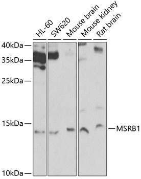
HL-60, SW620, Mouse brain, Mouse kidney, Rat brain
Cellular Localization:
Cytoplasm, Nucleus, Cytoskeleton.
Calculated MW:
13kDa
Observed MW:
13kDa
The protein encoded by this gene belongs to the methionine-R-sulfoxide reductase B (MsrB) family. Members of this family function as repair enzymes that protect proteins from oxidative stress by catalyzing the reduction of methionine-R-sulfoxides to methionines. This protein is highly expressed in liver and kidney, and is localized to the nucleus and cytosol. It is the only member of the MsrB family that is a selenoprotein, containing a selenocysteine (Sec) residue at its active site. It also has the highest methionine-R-sulfoxide reductase activity compared to other members containing cysteine in place of Sec. Sec is encoded by the UGA codon, which normally signals translation termination. The 3' UTRs of selenoprotein mRNAs contain a conserved stem-loop structure, designated the Sec insertion sequence (SECIS) element, that is necessary for the recognition of UGA as a Sec codon, rather than as a stop signal. A pseudogene of this locus has been identified on chromosome 19.
Purification Method
Affinity purification
Gene ID
51734
RRID
AB_2767321
Buffer Information
Store at -20℃. Avoid freeze / thaw cycles. Buffer: PBS containing 50% glycerol, preserved with proclin300 or sodium azide, pH 7.3.
Western blot analysis of various lysates using MSRB1 Rabbit pAb (CAB6737) at 1:1000 dilution. Secondary antibody: HRP-conjugated Goat anti-Rabbit IgG (H+L) (CABS014) at 1:10000 dilution. Lysates/proteins: 25μg per lane. Blocking buffer: 3% nonfat dry milk in TBST. Detection: ECL Basic Kit (AbGn00020). Exposure time: 30s.
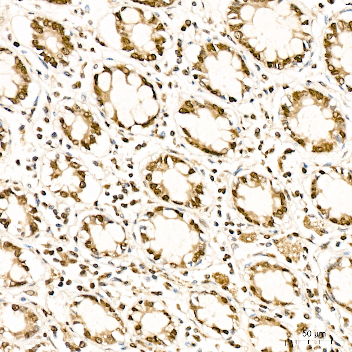
Immunohistochemistry analysis of paraffin-embedded Human colon tissue using MSRB1 Rabbit pAb (CAB6737) at a dilution of 1:300 (40x lens). High pressure antigen retrieval was performed with 0.01 M citrate buffer (pH 6.0) prior to IHC staining.
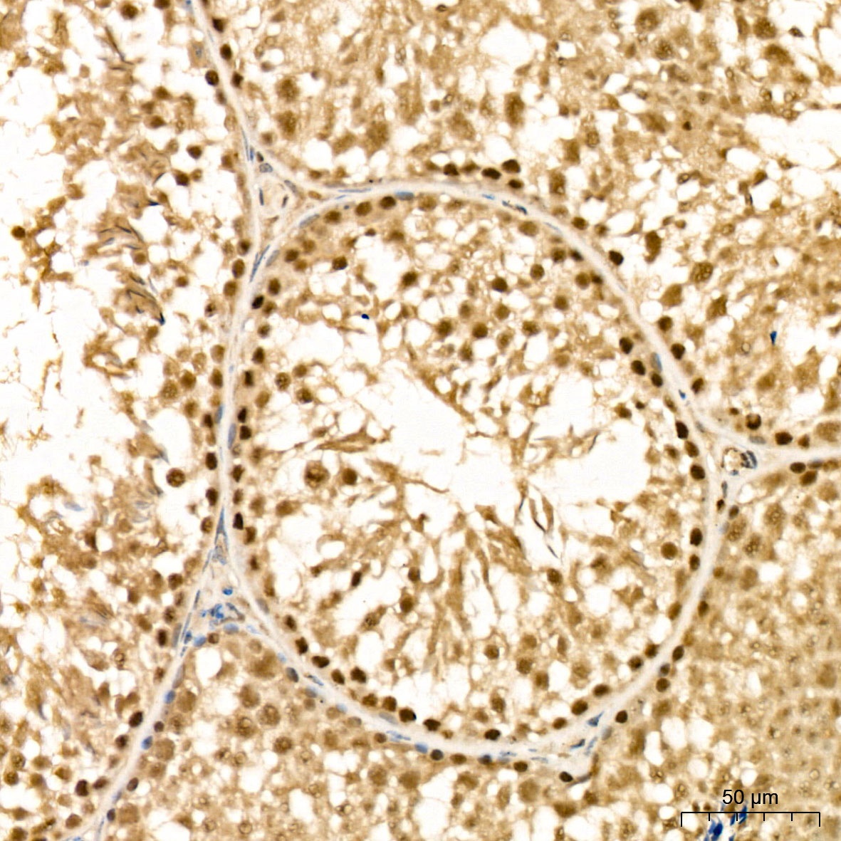
Immunohistochemistry analysis of paraffin-embedded Mouse testis tissue using MSRB1 Rabbit pAb (CAB6737) at a dilution of 1:300 (40x lens). High pressure antigen retrieval was performed with 0.01 M citrate buffer (pH 6.0) prior to IHC staining.
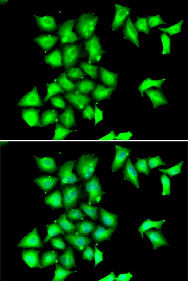
Immunofluorescence analysis of HeLa cells using MSRB1 Rabbit pAb (CAB6737). Secondary antibody: Cy3-conjugated Goat anti-Rabbit IgG (H+L) (CABS007) at 1:500 dilution. Blue: DAPI for nuclear staining.