The NSF Antibody (CAB0926) is a high-quality antibody developed for reliable detection and analysis of target proteins. This antibody, derived from rabbits, exhibits high reactivity with human samples and is suitable for use in Western blot applications. By binding to the NSF protein, it allows for the detection and analysis of this crucial molecule in a variety of cell types.NSF, or N-ethylmaleimide-sensitive factor, plays a vital role in the process of membrane fusion within cells, particularly in the context of vesicle trafficking and exocytosis. Dysregulation of NSF has been implicated in various diseases, including neurodegenerative disorders and viral infections.
This antibody is validated for use in WB, IF/ICC, IP, ELISA applications and has demonstrated reactivity against Human, Mouse, Rat samples.
Product Name:
NSF Antibody
SKU:
CAB0926
Size:
20μL, 100μL
Reactivity:
Human, Mouse, Rat
Conjugate:
Unconjugated
Immunogen:
Recombinant protein (or fragment).This information is considered to be commercially sensitive.
0.5μg-4μg antibody for 400μg-600μg extracts of whole cells
ELISA
Recommended starting concentration is 1 μg/mL. Please optimize the concentration based on your specific assay requirements.
Synonyms:
SKD2, DEE96, SEC18, NSF
Positive Sample:
PC-12, HepG2, C2C12, C6, SH-SY5Y, Mouse brain, Rat brain
Cellular Localization:
Cytoplasm.
Calculated MW:
83kDa
Observed MW:
80kDa
Enables PDZ domain binding activity and ionotropic glutamate receptor binding activity. Involved in intracellular protein transport; positive regulation of protein catabolic process; and positive regulation of receptor recycling. Located in Golgi apparatus; cytosol; and plasma membrane. Implicated in developmental and epileptic encephalopathy.
Purification Method
Affinity purification
Gene ID
4905
RRID
AB_2619672
Buffer Information
Store at -20℃. Avoid freeze / thaw cycles. Buffer: PBS containing 50% glycerol, preserved with proclin300 or sodium azide, pH 7.3.
Western blot analysis of various lysates using NSF Rabbit pAb (CAB0926) at 1:3000 dilution. Secondary antibody: HRP-conjugated Goat anti-Rabbit IgG (H+L) (CABS014) at 1:10000 dilution. Lysates/proteins: 25μg per lane. Blocking buffer: 3% nonfat dry milk in TBST. Detection: ECL Basic Kit (AbGn00020). Exposure time: 1s.
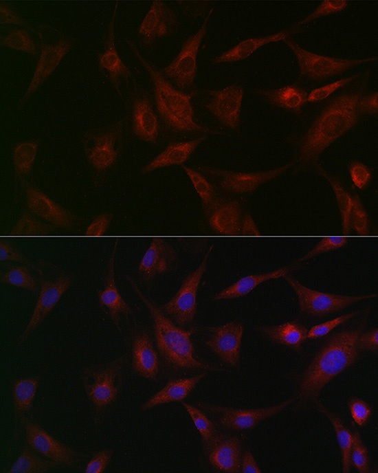
Immunofluorescence analysis of BALB-3T3 cells using NSF Rabbit pAb (CAB0926) at dilution of 1:100 (40x lens). Secondary antibody: Cy3-conjugated Goat anti-Rabbit IgG (H+L) (CABS007) at 1:500 dilution. Blue: DAPI for nuclear staining.
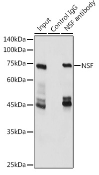
Immunoprecipitation analysis of 600 μg extracts of Mouse brain cells using 3 μg NSF antibody (CAB0926). Western blot was performed from the immunoprecipitate using NSF antibody (CAB0926) at a dilution of 1:3000.