The PCNA Antibody (CAB13336) is a high-quality antibody developed for reliable detection and analysis of target proteins. This antibody is raised in rabbits and exhibits high reactivity with human samples, making it a reliable option for Western blot applications.PCNA is a key player in maintaining genome stability and is essential for cell proliferation. Dysregulation of PCNA expression or function has been linked to various diseases, including cancer. By targeting PCNA with this antibody, researchers can analyze the protein's levels and localization in different cell types, providing insights into its role in cell cycle regulation and DNA damage response mechanisms.
This antibody is validated for use in WB, IHC-P, ELISA applications and has demonstrated reactivity against Human, Mouse, Rat samples.
Product Name:
PCNA Antibody
SKU:
CAB13336
Size:
20μL, 100μL
Reactivity:
Human, Mouse, Rat
Conjugate:
Unconjugated
Immunogen:
Synthetic peptide. This information is considered to be commercially sensitive.
Recommended starting concentration is 1 μg/mL. Please optimize the concentration based on your specific assay requirements.
Synonyms:
ATLD2, PCNA
Positive Sample:
HeLa, MCF7, 293T, C2C12, COS-7, Rat testis
Cellular Localization:
Nucleus.
Calculated MW:
29kDa
Observed MW:
34kDa
The protein encoded by this gene is found in the nucleus and is a cofactor of DNA polymerase delta. The encoded protein acts as a homotrimer and helps increase the processivity of leading strand synthesis during DNA replication. In response to DNA damage, this protein is ubiquitinated and is involved in the RAD6-dependent DNA repair pathway. Two transcript variants encoding the same protein have been found for this gene. Pseudogenes of this gene have been described on chromosome 4 and on the X chromosome.
Purification Method
Affinity purification
Gene ID
5111
RRID
AB_2760192
Buffer Information
Store at -20℃. Avoid freeze / thaw cycles. Buffer: PBS containing 50% glycerol, preserved with proclin300 or sodium azide, pH 7.3.
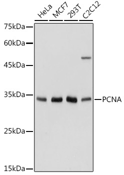
Western blot analysis of various lysates using PCNA Rabbit pAb (CAB13336) at 1:1000 dilution. Secondary antibody: HRP-conjugated Goat anti-Rabbit IgG (H+L) (CABS014) at 1:10000 dilution. Lysates/proteins: 25μg per lane. Blocking buffer: 3% nonfat dry milk in TBST. Detection: ECL Basic Kit (AbGn00020). Exposure time: 1s.
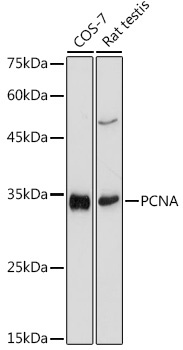
Western blot analysis of various lysates using PCNA Rabbit pAb (CAB13336) at 1:1000 dilution. Secondary antibody: HRP-conjugated Goat anti-Rabbit IgG (H+L) (CABS014) at 1:10000 dilution. Lysates/proteins: 25μg per lane. Blocking buffer: 3% nonfat dry milk in TBST. Detection: ECL Basic Kit (AbGn00020). Exposure time: 3s.
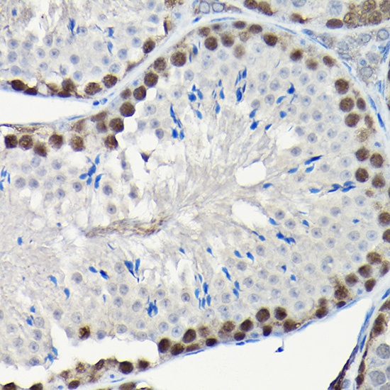
Immunohistochemistry analysis of paraffin-embedded Mouse testis using PCNA Rabbit pAb (CAB13336) at dilution of 1:200 (40x lens). High pressure antigen retrieval performed with 0.01M Citrate buffer (pH 6.0) prior to IHC staining.
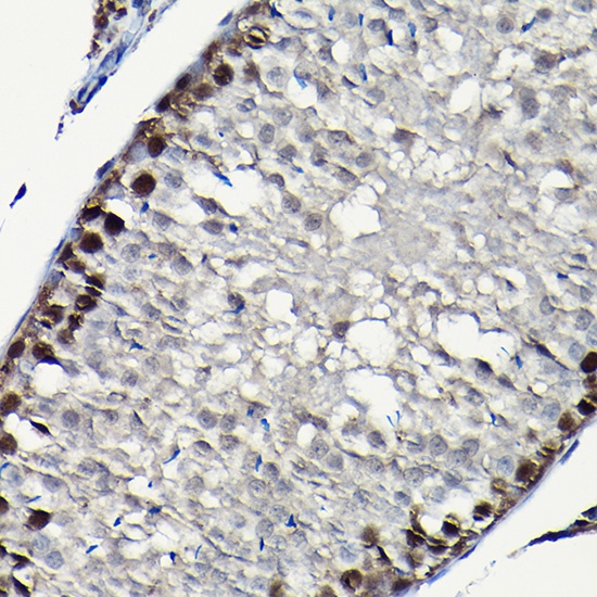
Immunohistochemistry analysis of paraffin-embedded Rat testis using PCNA Rabbit pAb (CAB13336) at dilution of 1:200 (40x lens). High pressure antigen retrieval performed with 0.01M Citrate buffer (pH 6.0) prior to IHC staining.