The PD-1/CD279 Antibody (CAB11973) is a high-quality antibody developed for reliable detection and analysis of target proteins. This cell surface receptor is a key regulator of immune responses, with its primary function being to inhibit T-cell activity and prevent autoimmune reactions.Raised in rabbits, this antibody is highly specific to human samples and has been validated for use in Western blot applications. By binding to the PDCD1 protein, it enables accurate detection and analysis, making it ideal for studies in immunology and cancer research.
This antibody is validated for use in WB, ELISA, IF-P applications and has demonstrated reactivity against Human, Mouse, Rat samples.
Product Name:
PD-1/CD279 Antibody
SKU:
CAB11973
Size:
20μL, 100μL
Reactivity:
Human, Mouse, Rat
Conjugate:
Unconjugated
Immunogen:
Recombinant protein (or fragment).This information is considered to be commercially sensitive.
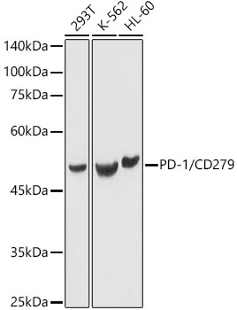
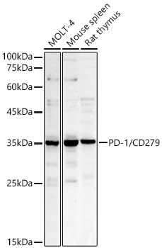
293T, K-562, HL-60, MOLT-4, Mouse spleen, Rat thymus
Cellular Localization:
Membrane, Single-Pass Type I Membrane Protein.
Calculated MW:
32kDa
Observed MW:
50kDa/35kDa
Programmed cell death protein 1 (PDCD1) is an immune-inhibitory receptor expressed in activated T cells; it is involved in the regulation of T-cell functions, including those of effector CD8+ T cells. In addition, this protein can also promote the differentiation of CD4+ T cells into T regulatory cells. PDCD1 is expressed in many types of tumors including melanomas, and has demonstrated to play a role in anti-tumor immunity. Moreover, this protein has been shown to be involved in safeguarding against autoimmunity, however, it can also contribute to the inhibition of effective anti-tumor and anti-microbial immunity.
Purification Method
Affinity purification
Gene ID
5133
RRID
AB_2758906
Buffer Information
Store at -20℃. Avoid freeze / thaw cycles. Buffer: PBS containing 50% glycerol, preserved with proclin300 or sodium azide, pH 7.3.
Western blot analysis of various lysates using PD-1/CD279 Rabbit pAb (CAB11973) at 1:1000 dilution. Secondary antibody: HRP-conjugated Goat anti-Rabbit IgG (H+L) (CABS014) at 1:10000 dilution. Lysates/proteins: 25μg per lane. Blocking buffer: 3% nonfat dry milk in TBST. Detection: ECL Basic Kit (AbGn00020). Exposure time: 1s.
Western blot analysis of various lysates using PD-1/CD279 Rabbit pAb (CAB11973) at 1:1000 dilution. Secondary antibody: HRP-conjugated Goat anti-Rabbit IgG (H+L) (CABS014) at 1:10000 dilution. Lysates / proteins: 25 μg per lane. Blocking buffer: 3 % nonfat dry milk in TBST. Detection: ECL Basic Kit (AbGn00020). Exposure time: 30s.


![Purified Anti-Human CD279/PD-1 Antibody [J110] (AGEL2401)](https://cdn11.bigcommerce.com/s-h68l9z2lnx/images/stencil/590x590/products/229804/602481/purified-anti-human-cd279pd-1-antibody-j110__05055.1706284862.jpg?c=2 "Purified Anti-Human CD279/PD-1 Antibody [J110] (AGEL2401)")