The Phospho-c-Myc-T58/S62 Monoclonal Antibody (CABP0988) is a high-quality antibody developed for reliable detection and analysis of target proteins. This antibody, developed using rabbit monoclonal technology, specifically targets the phosphorylation sites T58 and S62 on the c-Myc protein.This antibody is highly reactive with human samples, making it an excellent choice for experiments involving c-Myc signaling pathways. It has been validated for use in Western blot applications, enabling researchers to detect and analyze phosphorylated c-Myc in various cell types. The phosphorylation of c-Myc at T58 and S62 is known to have important implications in cancer research, as dysregulation of c-Myc signaling is commonly observed in many tumor types.
This antibody is validated for use in WB, ELISA applications and has demonstrated reactivity against Rat samples.
Product Name:
Phospho-c-Myc-T58/S62 Monoclonal Antibody
SKU:
CABP0988
Size:
20μL, 100μL
Reactivity:
Rat
Clone Number:
ARC1532
Conjugate:
Unconjugated
Immunogen:
Synthetic peptide. This information is considered to be commercially sensitive.
Sequence:
TPPL S
Tested Applications:
WBELISA
Recommended Dilution:
WB
1:500 - 1:2000
ELISA
Recommended starting concentration is 1 μg/mL. Please optimize the concentration based on your specific assay requirements.
Synonyms:
MRTL, MYCC, c-Myc, bHLHe39, Phospho-c-Myc-T58/S62
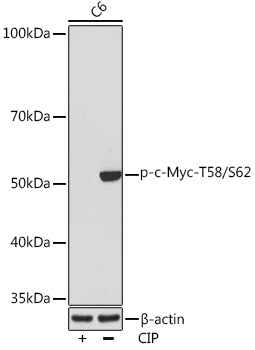
Positive Sample:
C6
Cellular Localization:
Nucleus, Nucleolus, Nucleoplasm.
Calculated MW:
51kDa
Observed MW:
57-65kDa
This gene is a proto-oncogene and encodes a nuclear phosphoprotein that plays a role in cell cycle progression, apoptosis and cellular transformation. The encoded protein forms a heterodimer with the related transcription factor MAX. This complex binds to the E box DNA consensus sequence and regulates the transcription of specific target genes. Amplification of this gene is frequently observed in numerous human cancers. Translocations involving this gene are associated with Burkitt lymphoma and multiple myeloma in human patients. There is evidence to show that translation initiates both from an upstream, in-frame non-AUG (CUG) and a downstream AUG start site, resulting in the production of two isoforms with distinct N-termini.
Purification Method
Affinity purification
Gene ID
4609
RRID
AB_2863882
Buffer Information
Store at -20℃. Avoid freeze / thaw cycles. Buffer: PBS containing 50% glycerol and 0.05% BSA, preserved with proclin300 or sodium azide, pH 7.3.
Western blot analysis of lysates from C6 cells, using Phospho-c-Myc-T58/S62 Rabbit mAb (CABP0988) at 1:1000 dilution. C6 cells were treated by CIP(20uL/400ul) at 37℃ for 1 hour. Secondary antibody: HRP-conjugated Goat anti-Rabbit IgG (H+L) (CABS014) at 1:10000 dilution. Lysates/proteins: 25μg per lane. Blocking buffer: 3% BSA. Detection: ECL Basic Kit (AbGn00020). Exposure time: 10s.