The Phospho-Myosin Light Chain 2-S19 Monoclonal Antibody (CABP1433) is a high-quality antibody developed for reliable detection and analysis of target proteins. This antibody, produced by Assay Genie, specifically targets the phosphorylation site at serine 19 of myosin light chain 2, allowing for precise detection and analysis of this post-translational modification in various experimental systems.This monoclonal antibody is highly sensitive and specific to phosphorylated myosin light chain 2 at serine 19, making it an essential tool for investigating the role of this protein in cellular processes such as muscle contraction, cell migration, and cytoskeletal dynamics.
This antibody is validated for use in WB, ELISA applications and has demonstrated reactivity against Human, Mouse, Rat samples.
HeLa treated with Calyculin A, C2C12 treated with Calyculin A
Cellular Localization:
Cell Cortex, Cytoplasm, Cytosol, Myofibril, Z Disc.
Calculated MW:
20kDa
Observed MW:
20kDa
Myosin, a structural component of muscle, consists of two heavy chains and four light chains. The protein encoded by this gene is a myosin light chain that may regulate muscle contraction by modulating the ATPase activity of myosin heads. The encoded protein binds calcium and is activated by myosin light chain kinase. Two transcript variants encoding different isoforms have been found for this gene.
Purification Method
Affinity purification
Gene ID
10398
Buffer Information
Store at -20℃. Avoid freeze / thaw cycles. Buffer: PBS containing 50% glycerol and 0.05% BSA, preserved with proclin300 or sodium azide, pH 7.3.
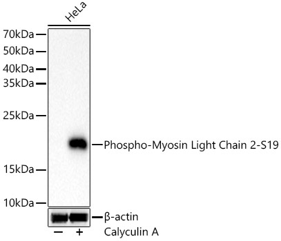
Western blot analysis of lysates from HeLa cells, using Phospho-Myosin Light Chain 2-S19 Rabbit mAb (CABP1433) at 1:1000 dilution. HeLa cells were treated with Calyculin A (100 nM) at 37℃ for 30 minutes after serum-starvation overnight. Secondary antibody: HRP-conjugated Goat anti-Rabbit IgG (H+L) (CABS014) at 1:10000 dilution. Lysates/proteins: 25μg per lane. Blocking buffer: 3% nonfat dry milk in TBST. Detection: ECL Basic Kit (AbGn00020). Exposure time: 10s.
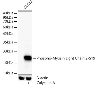
Western blot analysis of lysates from C2C12 cells, using Phospho-Myosin Light Chain 2-S19 Rabbit mAb (CABP1433) at 1:1000 dilution. C2C12 cells were treated with Calyculin A (100 nM) at 37℃ for 30 minutes after serum-starvation overnight. Secondary antibody: HRP-conjugated Goat anti-Rabbit IgG (H+L) (CABS014) at 1:10000 dilution. Lysates/proteins: 25μg per lane. Blocking buffer: 3% nonfat dry milk in TBST. Detection: ECL Basic Kit (AbGn00020). Exposure time: 15s.
 at 1:1000 dilution. HeLa cells were treated by Calyculin A (100 nM) at 37℃ for 30 minutes after serum-starvation overnight. Secondary antibody: HRP Goat Anti-Rabbit IgG (H+L) at 1:10000 dilution. Lysates/proteins: 25μg per lane. Blocking buffer: 3% nonfat dry milk in TBST.")
 at 1:1000 dilution. HeLa cells were treated by Calyculin A (100 nM) at 37℃ for 30 minutes after serum-starvation overnight. Secondary antibody: HRP Goat Anti-Rabbit IgG (H+L) at 1:10000 dilution. Lysates/proteins: 25μg per lane. Blocking buffer: 3% nonfat dry milk in TBST.")


