The Phospho-p53-S9 Antibody (CABP0085) is a high-quality antibody developed for reliable detection and analysis of target proteins. This antibody, produced in rabbits, exhibits high specificity and sensitivity towards human samples, making it an ideal choice for Western blot analysis.Phosphorylation of p53 at Serine 9 is known to play a crucial role in its activation and downstream signaling pathways. By targeting this specific phosphorylation site, researchers can gain valuable insights into the mechanisms governing p53 activation and its impact on cell cycle regulation, apoptosis, and DNA repair processes.The Phospho-p53 (S9) Polyclonal Antibody is a valuable asset for studies in cancer biology, as dysregulation of the p53 pathway is a common feature in many types of cancer.
This antibody is validated for use in WB, IP, ELISA applications and has demonstrated reactivity against Human samples.
Product Name:
Phospho-p53-S9 Antibody
SKU:
CABP0085
Size:
20μL, 100μL
Reactivity:
Human
Conjugate:
Unconjugated
Immunogen:
Synthetic peptide. This information is considered to be commercially sensitive.
Sequence:
DPSV E
Tested Applications:
WBIPELISA
Recommended Dilution:
WB
1:500 - 1:2000
IP
0.5μg-4μg antibody for 200μg-400μg extracts of whole cells
ELISA
Recommended starting concentration is 1 μg/mL. Please optimize the concentration based on your specific assay requirements.
This gene encodes a tumor suppressor protein containing transcriptional activation, DNA binding, and oligomerization domains. The encoded protein responds to diverse cellular stresses to regulate expression of target genes, thereby inducing cell cycle arrest, apoptosis, senescence, DNA repair, or changes in metabolism. Mutations in this gene are associated with a variety of human cancers, including hereditary cancers such as Li-Fraumeni syndrome. Alternative splicing of this gene and the use of alternate promoters result in multiple transcript variants and isoforms. Additional isoforms have also been shown to result from the use of alternate translation initiation codons from identical transcript variants (PMIDs: 12032546, 20937277).
Purification Method
Affinity purification
Gene ID
7157
RRID
AB_2771382
Buffer Information
Store at -20℃. Avoid freeze / thaw cycles. Buffer: PBS containing 50% glycerol, preserved with proclin300 or sodium azide, pH 7.3.
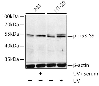
Western blot analysis of lysates from 293 and HT-29 cells, using Phospho-p53-S9 Rabbit pAb (CABP0085) at 1:1000 dilution. 293 cells were treated with UV for 15-30 minutes. HT-29 cells were treated with UV for 15-30 minutes. Secondary antibody: HRP-conjugated Goat anti-Rabbit IgG (H+L) (CABS014) at 1:10000 dilution. Lysates/proteins: 25μg per lane. Blocking buffer: 3% BSA. Detection: ECL Basic Kit (AbGn00020). Exposure time: 3min.
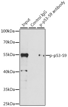
Immunoprecipitation analysis of 200 μg extracts of 293T cells, using 3 μg Phospho-p53-S9 pAb (CABP0085). Western blot was performed from the immunoprecipitate using Phospho-p53-S9 pAb (CABP0085) at a dilution of 1:1000. 293T cells were treated with UV at room temperature for 30 minutes after serum-starvation overnight, and then treated with 10% FBS at 37℃ for 30 minutes.