The PLA2G2D Antibody (CAB7165) is a high-quality antibody developed for reliable detection and analysis of target proteins. This antibody, produced in rabbits, exhibits high specificity and reactivity towards human samples, making it an excellent choice for Western blot experiments. By targeting the PLA2G2D protein, researchers can effectively study its expression and function in various biological samples, providing insights into its role in lipid signaling pathways and inflammatory responses.PLA2G2D, also known as phospholipase A2 group IID, is known to play a crucial role in lipid metabolism by catalyzing the hydrolysis of membrane phospholipids to release fatty acids and bioactive lipid mediators.
This antibody is validated for use in WB, IHC-P, IF/ICC, ELISA applications and has demonstrated reactivity against Human, Rat samples.
Product Name:
PLA2G2D Antibody
SKU:
CAB7165
Size:
20μL, 100μL
Reactivity:
Human, Rat
Conjugate:
Unconjugated
Immunogen:
Recombinant protein (or fragment).This information is considered to be commercially sensitive.
Recommended starting concentration is 1 μg/mL. Please optimize the concentration based on your specific assay requirements.
Synonyms:
SPLASH, sPLA2S, PLA2IID, sPLA2-IID, PLA2G2D
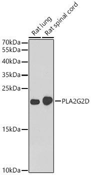
Positive Sample:
Daudi, RPMI 8226, Rat lung, Rat spinal cord
Cellular Localization:
Secreted.
Calculated MW:
17kDa
Observed MW:
22kDa
This gene encodes a secreted member of the phospholipase A2 family, and is found in a cluster of related family members on chromosome 1. Phospholipase A2 family members hydrolyze the sn-2 fatty acid ester bond of glycerophospholipids to produce lysophospholipids and free fatty acid. This gene may be involved in inflammation and immune response, and in weight loss associated with chronic obstructive pulmonary disease. Alternative splicing results in multiple transcript variants encoding different isoforms.
Purification Method
Affinity purification
Gene ID
26279
RRID
AB_2767717
Buffer Information
Store at -20℃. Avoid freeze / thaw cycles. Buffer: PBS containing 50% glycerol, preserved with proclin300 or sodium azide, pH 7.3.
Western blot analysis of various lysates using PLA2G2D Rabbit pAb (CAB7165) at 1:1000 dilution. Secondary antibody: HRP-conjugated Goat anti-Rabbit IgG (H+L) (CABS014) at 1:10000 dilution. Lysates / proteins: 25 μg per lane. Blocking buffer: 3 % nonfat dry milk in TBST. Detection: ECL Basic Kit (AbGn00020). Exposure time: 90s.
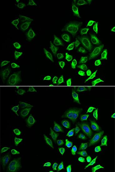
Immunofluorescence analysis of U2OS cells using PLA2G2D Rabbit pAb (CAB7165). Secondary antibody: Cy3-conjugated Goat anti-Rabbit IgG (H+L) (CABS007) at 1:500 dilution. Blue: DAPI for nuclear staining.