The RPA70 Antibody (CAB9841) is a high-quality antibody developed for reliable detection and analysis of target proteins. This antibody, produced in rabbits, exhibits high specificity and reactivity towards human samples, making it a reliable choice for various research applications, particularly in Western blot analyses.RPE65 is a vital enzyme found in the retina that plays a crucial role in the conversion of vitamin A into the visual pigment rhodopsin, enabling the process of light detection by photoreceptor cells. Mutations in the RPE65 gene can lead to retinal degenerative disorders such as Leber congenital amaurosis and retinitis pigmentosa, highlighting the importance of studying this protein for potential therapeutic interventions.
This antibody is validated for use in WB, ELISA, IF-P applications and has demonstrated reactivity against Human, Mouse, Rat samples.
Product Name:
RPA70 Antibody
SKU:
CAB9841
Size:
20μL, 100μL
Reactivity:
Human, Mouse, Rat
Conjugate:
Unconjugated
Immunogen:
Recombinant protein (or fragment).This information is considered to be commercially sensitive.
The protein encoded by this gene is a component of the vitamin A visual cycle of the retina which supplies the 11-cis retinal chromophore of the photoreceptors opsin visual pigments. It is a member of the carotenoid cleavage oxygenase superfamily. All members of this superfamily are non-heme iron oxygenases with a seven-bladed propeller fold and oxidatively cleave carotenoid carbon:carbon double bonds. However, the protein encoded by this gene has acquired a divergent function that involves the concerted O-alkyl ester cleavage of its all-trans retinyl ester substrate and all-trans to 11-cis double bond isomerization of the retinyl moiety. As such, it performs the essential enzymatic isomerization step in the synthesis of 11-cis retinal. Mutations in this gene are associated with early-onset severe blinding disorders such as Leber congenital.
Purification Method
Affinity purification
Gene ID
6121
RRID
AB_2772068
Buffer Information
Store at -20℃. Avoid freeze / thaw cycles. Buffer: PBS containing 50% glycerol, preserved with proclin300 or sodium azide, pH 7.3.
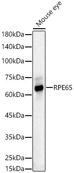
Western blot analysis of lysates from Mouse eye, using RPE65 Rabbit pAb (CAB9841) at 1:3000 dilution. Secondary antibody: HRP-conjugated Goat anti-Rabbit IgG (H+L) (CABS014) at 1:10000 dilution. Lysates/proteins: 25μg per lane. Blocking buffer: 3% nonfat dry milk in TBST. Detection: ECL Basic Kit (AbGn00020). Exposure time: 30s.
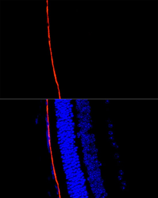
Perform microwave antigen retrieval with 10 mM citrate buffer pH 6.0 before commencing with IF staining protocol.Immunofluorescence analysis of paraffin-embedded mouse retina using RPE65 Rabbit pAb (CAB9841) at dilution of 1:300 (40x lens). Secondary antibody: Cy3-conjugated Goat anti-Rabbit IgG (H+L) (CABS007) at 1:500 dilution. Blue: DAPI for nuclear staining.