The RPA70 Monoclonal Antibody (CAB9615) is a high-quality antibody developed for reliable detection and analysis of target proteins. This antibody is specifically designed to target RPE65 in human samples, making it ideal for Western blot applications. By binding to the RPE65 protein, this antibody allows for accurate detection and analysis in various cell types, facilitating research in ophthalmology and vision science.RPE65 is essential for the conversion of retinyl esters to 11-cis-retinol in the visual cycle, a crucial process for maintaining proper vision.
This antibody is validated for use in WB, IHC-P, ELISA, IF-P applications and has demonstrated reactivity against Mouse, Rat samples.
Product Name:
RPA70 Monoclonal Antibody
SKU:
CAB9615
Size:
20μL, 100μL
Reactivity:
Mouse, Rat
Clone Number:
ARC1659
Conjugate:
Unconjugated
Immunogen:
Synthetic peptide. This information is considered to be commercially sensitive.
The protein encoded by this gene is a component of the vitamin A visual cycle of the retina which supplies the 11-cis retinal chromophore of the photoreceptors opsin visual pigments. It is a member of the carotenoid cleavage oxygenase superfamily. All members of this superfamily are non-heme iron oxygenases with a seven-bladed propeller fold and oxidatively cleave carotenoid carbon:carbon double bonds. However, the protein encoded by this gene has acquired a divergent function that involves the concerted O-alkyl ester cleavage of its all-trans retinyl ester substrate and all-trans to 11-cis double bond isomerization of the retinyl moiety. As such, it performs the essential enzymatic isomerization step in the synthesis of 11-cis retinal. Mutations in this gene are associated with early-onset severe blinding disorders such as Leber congenital.
Purification Method
Affinity purification
Gene ID
6121
RRID
AB_2863738
Buffer Information
Store at -20℃. Avoid freeze / thaw cycles. Buffer: PBS containing 50% glycerol and 0.05% BSA, preserved with proclin300 or sodium azide, pH 7.3.
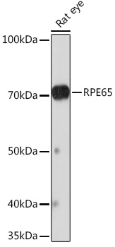
Western blot analysis of lysates from Rat eye, using RPE65 Rabbit mAb (CAB9615) at 1:1000 dilution. Secondary antibody: HRP-conjugated Goat anti-Rabbit IgG (H+L) (CABS014) at 1:10000 dilution. Lysates/proteins: 25μg per lane. Blocking buffer: 3% nonfat dry milk in TBST. Detection: ECL Basic Kit (AbGn00020). Exposure time: 3min.
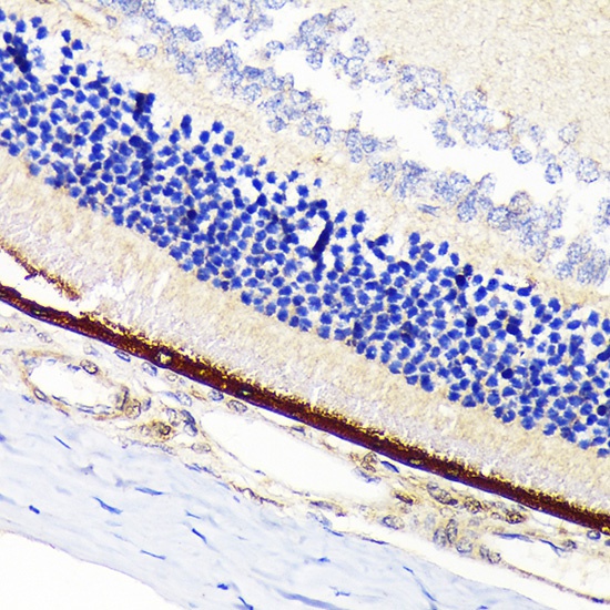
Immunohistochemistry analysis of paraffin-embedded Rat retina using RPE65 Rabbit mAb (CAB9615) at dilution of 1:100 (40x lens). Microwave antigen retrieval performed with 0.01M Tris/EDTA Buffer (pH 9.0) prior to IHC staining.
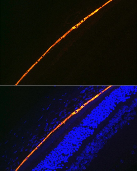
Immunofluorescence analysis of paraffin-embedded rat eye using RPE65 Rabbit mAb (CAB9615) at dilution of 1:100 (40x lens). Secondary antibody: Cy3-conjugated Goat anti-Rabbit IgG (H+L) (CABS007) at 1:500 dilution. Blue: DAPI for nuclear staining.
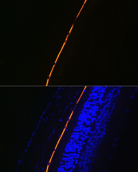
Immunofluorescence analysis of paraffin-embedded mouse eye using RPE65 Rabbit mAb (CAB9615) at dilution of 1:100 (40x lens). Secondary antibody: Cy3-conjugated Goat anti-Rabbit IgG (H+L) (CABS007) at 1:500 dilution. Blue: DAPI for nuclear staining.