The RPS3 Antibody (CAB2533) is a high-quality antibody developed for reliable detection and analysis of target proteins. This antibody, produced in rabbits, is highly specific to human RPS3 and has been validated for use in Western blot applications. By binding to the RPS3 protein, this antibody facilitates the detection and analysis of RPS3 expression in various cell types, making it ideal for research in molecular biology and cancer biology.RPS3, a multifunctional protein, is known to play a crucial role in ribosome biogenesis and translation initiation, making it essential for cell growth and proliferation.
This antibody is validated for use in WB, IHC-P, IF/ICC, ELISA applications and has demonstrated reactivity against Human, Mouse, Rat samples.
Product Name:
RPS3 Antibody
SKU:
CAB2533
Size:
20μL, 100μL
Reactivity:
Human, Mouse, Rat
Conjugate:
Unconjugated
Immunogen:
Recombinant protein (or fragment).This information is considered to be commercially sensitive.
Ribosomes, the organelles that catalyze protein synthesis, consist of a small 40S subunit and a large 60S subunit. Together these subunits are composed of 4 RNA species and approximately 80 structurally distinct proteins. This gene encodes a ribosomal protein that is a component of the 40S subunit, where it forms part of the domain where translation is initiated. The protein belongs to the S3P family of ribosomal proteins. Studies of the mouse and rat proteins have demonstrated that the protein has an extraribosomal role as an endonuclease involved in the repair of UV-induced DNA damage. The protein appears to be located in both the cytoplasm and nucleus but not in the nucleolus. Higher levels of expression of this gene in colon adenocarcinomas and adenomatous polyps compared to adjacent normal colonic mucosa have been observed. This gene is co-transcribed with the small nucleolar RNA genes U15A and U15B, which are located in its first and fifth introns, respectively. As is typical for genes encoding ribosomal proteins, there are multiple processed pseudogenes of this gene dispersed through the genome. Multiple alternatively spliced transcript variants encoding different isoforms have been found for this gene.
Purification Method
Affinity purification
Gene ID
6188
RRID
AB_2764424
Buffer Information
Store at -20℃. Avoid freeze / thaw cycles. Buffer: PBS containing 50% glycerol, preserved with proclin300 or sodium azide, pH 7.3.
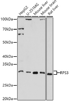
Western blot analysis of various lysates using RPS3 Rabbit pAb (CAB2533) at 1:1000 dilution. Secondary antibody: HRP-conjugated Goat anti-Rabbit IgG (H+L) (CABS014) at 1:10000 dilution. Lysates/proteins: 25μg per lane. Blocking buffer: 3% nonfat dry milk in TBST. Detection: ECL Basic Kit (AbGn00020). Exposure time: 180s.
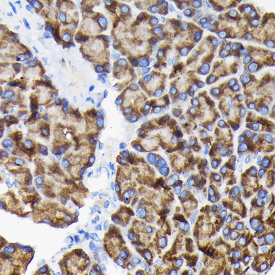
Immunohistochemistry analysis of paraffin-embedded Rat pancreas using RPS3 Rabbit pAb (CAB2533) at dilution of 1:100 (40x lens). Microwave antigen retrieval performed with 0.01M Tris/EDTA Buffer (pH 9.0) prior to IHC staining.
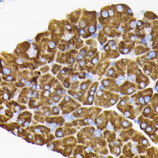
Immunohistochemistry analysis of paraffin-embedded Mouse pancreas using RPS3 Rabbit pAb (CAB2533) at dilution of 1:100 (40x lens). Microwave antigen retrieval performed with 0.01M Tris/EDTA Buffer (pH 9.0) prior to IHC staining.
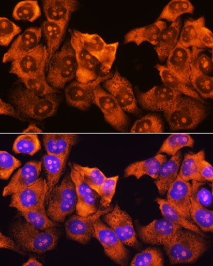
Immunofluorescence analysis of HeLa cells using RPS3 Rabbit pAb (CAB2533) at dilution of 1:100. Secondary antibody: Cy3-conjugated Goat anti-Rabbit IgG (H+L) (CABS007) at 1:500 dilution. Blue: DAPI for nuclear staining.
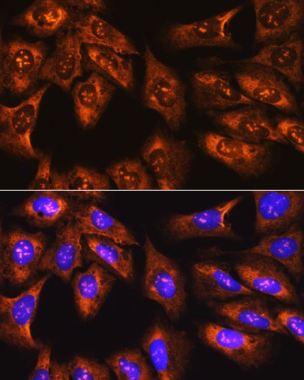
Immunofluorescence analysis of U-2 OS cells using RPS3 Rabbit pAb (CAB2533) at dilution of 1:100. Secondary antibody: Cy3-conjugated Goat anti-Rabbit IgG (H+L) (CABS007) at 1:500 dilution. Blue: DAPI for nuclear staining.