The SMPD4 Antibody (CAB15473) is a high-quality antibody developed for reliable detection and analysis of target proteins. The antibody, produced in rabbits, has high specificity for human samples and is validated for use in Western blot applications.SMPD4 is known to play a crucial role in cellular processes such as apoptosis, cell growth, and differentiation, making it a potential therapeutic target for various diseases, including cancer and neurodegenerative disorders. By detecting and analyzing SMPD4 protein levels in different cell types, this antibody enables researchers to investigate the function and regulation of SMPD4 in various biological contexts.
This antibody is validated for use in WB, IF/ICC, ELISA applications and has demonstrated reactivity against Human, Mouse samples.
Product Name:
SMPD4 Antibody
SKU:
CAB15473
Size:
20μL, 100μL
Reactivity:
Human, Mouse
Conjugate:
Unconjugated
Immunogen:
Recombinant protein (or fragment).This information is considered to be commercially sensitive.
The protein encoded by this gene is a sphingomyelinase that catalyzes the hydrolysis of membrane sphingomyelin to form phosphorylcholine and ceramide. This gene is activated by DNA damage, cellular stress, and tumor necrosis factor, but it is downregulated by wild-type p53. The encoded protein localizes to the endoplasmic reticulum and Golgi network.
Purification Method
Affinity purification
Gene ID
55627
RRID
AB_2762871
Buffer Information
Store at -20℃. Avoid freeze / thaw cycles. Buffer: PBS with 0.01% thimerosal,50% glycerol,pH7.3.
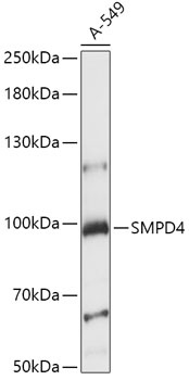
Western blot analysis of lysates from A-549 cells, using SMPD4 Rabbit pAb (CAB15473) at 1:1000 dilution. Secondary antibody: HRP-conjugated Goat anti-Rabbit IgG (H+L) (CABS014) at 1:10000 dilution. Lysates/proteins: 25μg per lane. Blocking buffer: 3% nonfat dry milk in TBST. Detection: ECL Basic Kit (AbGn00020). Exposure time: 15s.
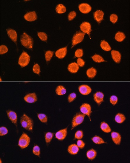
Immunofluorescence analysis of L929 cells using SMPD4 Rabbit pAb (CAB15473) at dilution of 1:100. Secondary antibody: Cy3-conjugated Goat anti-Rabbit IgG (H+L) (CABS007) at 1:500 dilution. Blue: DAPI for nuclear staining.