The ST3GAL3 Antibody (CAB6753) is a high-quality antibody developed for reliable detection and analysis of target proteins. This antibody, produced in rabbits, exhibits high reactivity with human samples and has been validated for use in Western blot applications. By specifically binding to the ST3GAL3 protein, this antibody enables accurate detection and analysis in a variety of cell types, making it an ideal choice for studies in glycobiology and cancer research.
This antibody is validated for use in WB, IHC-P, IF/ICC, ELISA applications and has demonstrated reactivity against Human, Mouse, Rat samples.
Product Name:
ST3GAL3 Antibody
SKU:
CAB6753
Size:
20μL, 100μL
Reactivity:
Human, Mouse, Rat
Conjugate:
Unconjugated
Immunogen:
Recombinant protein (or fragment).This information is considered to be commercially sensitive.
Recommended starting concentration is 1 μg/mL. Please optimize the concentration based on your specific assay requirements.
Synonyms:
ST3N, DEE15, MRT12, SIAT6, EIEE15, ST3GALII, ST3GalIII, ST3Gal III, ST3GAL3
Positive Sample:
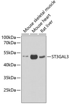
Mouse skeletal muscle, Mouse heart, Rat liver
Cellular Localization:
Golgi Apparatus, Golgi Stack Membrane, Secreted, Single-Pass Type Ii Membrane Protein.
Calculated MW:
42kDa
Observed MW:
42kDa
The protein encoded by this gene is a type II membrane protein that catalyzes the transfer of sialic acid from CMP-sialic acid to galactose-containing substrates. The encoded protein is normally found in the Golgi apparatus but can be proteolytically processed to a soluble form. This protein is a member of glycosyltransferase family 29. Mutations in this gene have been associated with a form of autosomal recessive nonsymdromic cognitive disability as well as infantile epileptic encephalopathy. Multiple transcript variants encoding several different isoforms have been found for this gene.
Purification Method
Affinity purification
Gene ID
6487
RRID
AB_2767337
Buffer Information
Store at -20℃. Avoid freeze / thaw cycles. Buffer: PBS containing 50% glycerol, preserved with proclin300 or sodium azide, pH 7.3.
Western blot analysis of various lysates using ST3GAL3 Rabbit pAb (CAB6753) at 1:1000 dilution. Secondary antibody: HRP-conjugated Goat anti-Rabbit IgG (H+L) (CABS014) at 1:10000 dilution. Lysates/proteins: 25μg per lane. Blocking buffer: 3% nonfat dry milk in TBST. Detection: ECL Basic Kit (AbGn00020). Exposure time: 90s.
Immunohistochemistry analysis of paraffin-embedded Human liver damage using ST3GAL3 Rabbit pAb (CAB6753) at dilution of 1:100 (40x lens). Microwave antigen retrieval performed with 0.01M PBS Buffer (pH 7.2) prior to IHC staining.
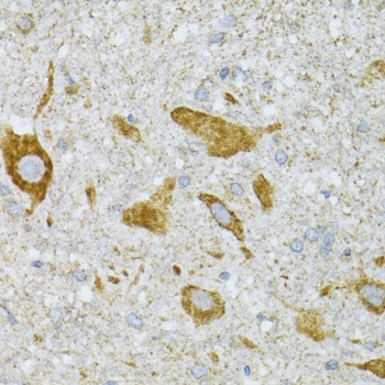
Immunohistochemistry analysis of paraffin-embedded Mouse spinal cord using ST3GAL3 Rabbit pAb (CAB6753) at dilution of 1:100 (40x lens). Microwave antigen retrieval performed with 0.01M PBS Buffer (pH 7.2) prior to IHC staining.
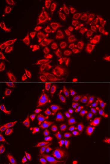
Immunofluorescence analysis of HeLa cells using ST3GAL3 Rabbit pAb (CAB6753). Secondary antibody: Cy3-conjugated Goat anti-Rabbit IgG (H+L) (CABS007) at 1:500 dilution. Blue: DAPI for nuclear staining.