The TGM1 Antibody (CAB13359) is a high-quality antibody developed for reliable detection and analysis of target proteins. This antibody, raised in rabbits, is highly specific to human TGM1 and has been validated for use in Western blot and immunohistochemistry applications.TGM1 is crucial for normal skin development and barrier function, making it a key target for investigation in dermatological research. Mutations in the TGM1 gene are associated with several skin disorders, including lamellar ichthyosis and other forms of congenital ichthyosis.
This antibody is validated for use in WB, ELISA applications and has demonstrated reactivity against Human, Mouse, Rat samples.
Product Name:
TGM1 Antibody
SKU:
CAB13359
Size:
20μL, 100μL
Reactivity:
Human, Mouse, Rat
Conjugate:
Unconjugated
Immunogen:
Recombinant protein (or fragment).This information is considered to be commercially sensitive.
Recommended starting concentration is 1 μg/mL. Please optimize the concentration based on your specific assay requirements.
Synonyms:
LI, KTG, LI1, TGK, ICR2, ARCI1, TGASE, TGM1
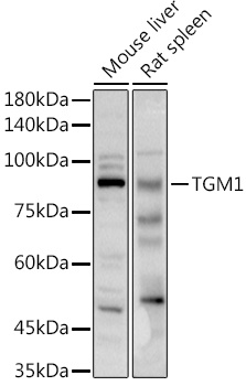
Positive Sample:
Mouse liver, Rat spleen
Cellular Localization:
Lipid-Anchor, Membrane.
Calculated MW:
90kDa
Observed MW:
90kDa
The protein encoded by this gene is a membrane protein that catalyzes the addition of an alkyl group from an akylamine to a glutamine residue of a protein, forming an alkylglutamine in the protein. This protein alkylation leads to crosslinking of proteins and catenation of polyamines to proteins. This gene contains either one or two copies of a 22 nt repeat unit in its 3' UTR. Mutations in this gene have been associated with autosomal recessive lamellar ichthyosis (LI) and nonbullous congenital ichthyosiform erythroderma (NCIE).
Purification Method
Affinity purification
Gene ID
7051
RRID
AB_2760216
Buffer Information
Store at -20℃. Avoid freeze / thaw cycles. Buffer: PBS containing 50% glycerol, preserved with proclin300 or sodium azide, pH 7.3.
Western blot analysis of various lysates using TGM1 Rabbit pAb (CAB13359) at 1:1000 dilution. Secondary antibody: HRP-conjugated Goat anti-Rabbit IgG (H+L) (CABS014) at 1:10000 dilution. Lysates/proteins: 25μg per lane. Blocking buffer: 3% nonfat dry milk in TBST. Detection: ECL Basic Kit (AbGn00020). Exposure time: 90s.