The TP73 Antibody (CAB0385) is a high-quality antibody developed for reliable detection and analysis of target proteins. This antibody, produced using rabbits, exhibits high reactivity with human samples and has been validated for use in applications like Western blotting. By specifically binding to the TP73 protein, this antibody allows for precise detection and analysis in a variety of cell types, making it an excellent choice for studies in cancer biology and molecular biology.TP73, a member of the p53 family of tumor suppressor proteins, plays a crucial role in controlling cell cycle progression and inducing programmed cell death.
This antibody is validated for use in WB, IHC-P, ELISA, IF-P applications and has demonstrated reactivity against Human, Mouse samples.
Product Name:
TP73 Antibody
SKU:
CAB0385
Size:
20μL, 100μL
Reactivity:
Human, Mouse
Conjugate:
Unconjugated
Immunogen:
Recombinant protein (or fragment).This information is considered to be commercially sensitive.
Recommended starting concentration is 1 μg/mL. Please optimize the concentration based on your specific assay requirements.
Synonyms:
P73, CILD47, TP73
Positive Sample:
293T
Cellular Localization:
Cytoplasm, Nucleus.
Calculated MW:
70kDa
Observed MW:
80kDa
This gene encodes a member of the p53 family of transcription factors involved in cellular responses to stress and development. It maps to a region on chromosome 1p36 that is frequently deleted in neuroblastoma and other tumors, and thought to contain multiple tumor suppressor genes. The demonstration that this gene is monoallelically expressed (likely from the maternal allele), supports the notion that it is a candidate gene for neuroblastoma. Many transcript variants resulting from alternative splicing and/or use of alternate promoters have been found for this gene, but the biological validity and the full-length nature of some variants have not been determined.
Purification Method
Affinity purification
Gene ID
7161
RRID
AB_2757163
Buffer Information
Store at -20℃. Avoid freeze / thaw cycles. Buffer: PBS with 0.01% thimerosal,50% glycerol,pH7.3.
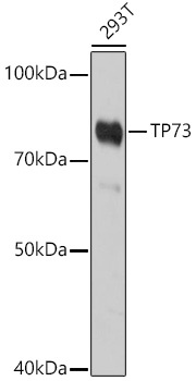
Western blot analysis of lysates from 293T cells, using TP73 Rabbit pAb (CAB0385) at 1:1000 dilution. Secondary antibody: HRP-conjugated Goat anti-Rabbit IgG (H+L) (CABS014) at 1:10000 dilution. Lysates/proteins: 25μg per lane. Blocking buffer: 3% nonfat dry milk in TBST. Detection: ECL Basic Kit (AbGn00020). Exposure time: 180s.
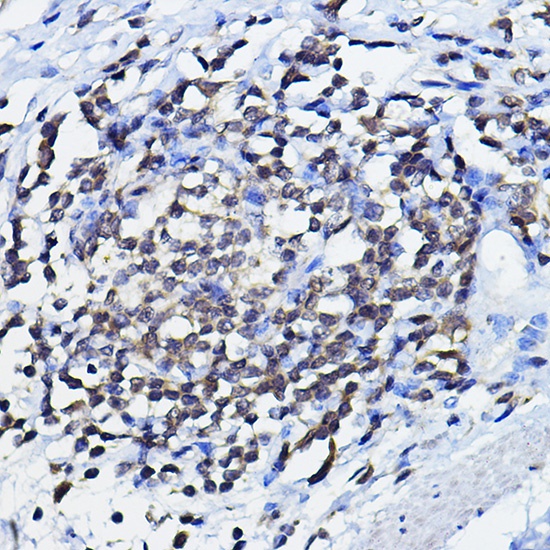
Immunohistochemistry analysis of paraffin-embedded Human lymph node using TP73 Rabbit pAb (CAB0385) at dilution of 1:100 (40x lens). High pressure antigen retrieval performed with 0.01M Citrate buffer (pH 6.0) prior to IHC staining.
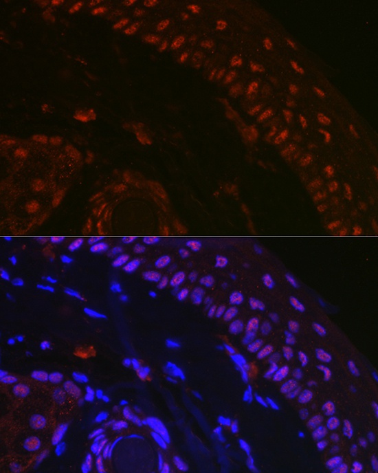
Immunofluorescence analysis of paraffin-embedded mouse skin using TP73 Rabbit pAb (CAB0385) at dilution of 1:100 (40x lens). Secondary antibody: Cy3-conjugated Goat anti-Rabbit IgG (H+L) (CABS007) at 1:500 dilution. Blue: DAPI for nuclear staining.