The TRAF7 Antibody (CAB3095) is a high-quality antibody developed for reliable detection and analysis of target proteins. This antibody, produced in rabbits, has high reactivity with human samples and is validated for use in Western blot applications. By specifically binding to TRAF7 protein, this antibody enables accurate detection and analysis in a wide range of cell types, making it an essential component for studies in cell signaling pathways and developmental biology.TRAF7, also known as TNF receptor-associated factor 7, is a crucial regulator of diverse cellular functions, including cell survival, proliferation, and immune response modulation.
This antibody is validated for use in WB, IHC-P, IF/ICC, ELISA applications and has demonstrated reactivity against Human, Mouse, Rat samples.
Product Name:
TRAF7 Antibody
SKU:
CAB3095
Size:
20μL, 100μL
Reactivity:
Human, Mouse, Rat
Conjugate:
Unconjugated
Immunogen:
Recombinant protein (or fragment).This information is considered to be commercially sensitive.
Recommended starting concentration is 1 μg/mL. Please optimize the concentration based on your specific assay requirements.
Synonyms:
RFWD1, RNF119, CAFDADD, TRAF7
Positive Sample:
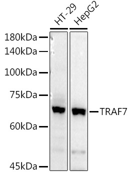
HT-29, HepG2
Cellular Localization:
Cytoplasmic Vesicle.
Calculated MW:
75kDa
Observed MW:
67kDa
Tumor necrosis factor (TNF; see MIM 191160) receptor-associated factors, such as TRAF7, are signal transducers for members of the TNF receptor superfamily (see MIM 191190). TRAFs are composed of an N-terminal cysteine/histidine-rich region containing zinc RING and/or zinc finger motifs; a coiled-coil (leucine zipper) motif; and a homologous region that defines the TRAF family, the TRAF domain, which is involved in self-association and receptor binding.
Purification Method
Affinity purification
Gene ID
84231
RRID
AB_2764896
Buffer Information
Store at -20℃. Avoid freeze / thaw cycles. Buffer: PBS containing 50% glycerol, preserved with proclin300 or sodium azide, pH 7.3.
Western blot analysis of various lysates using TRAF7 Rabbit pAb (CAB3095) at 1:1000 dilution. Secondary antibody: HRP-conjugated Goat anti-Rabbit IgG (H+L) (CABS014) at 1:10000 dilution. Lysates/proteins: 25μg per lane. Blocking buffer: 3% nonfat dry milk in TBST. Detection: ECL Basic Kit (AbGn00020). Exposure time: 180s.
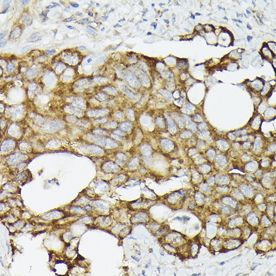
Immunohistochemistry analysis of paraffin-embedded Human colon carcinoma using TRAF7 Rabbit pAb (CAB3095) at dilution of 1:50 (40x lens). High pressure antigen retrieval performed with 0.01M Citrate buffer (pH 6.0) prior to IHC staining.
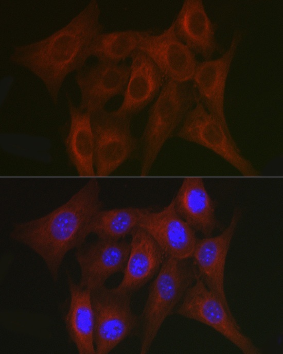
Immunofluorescence analysis of NIH/3T3 cells using TRAF7 Rabbit pAb (CAB3095) at dilution of 1:50. Secondary antibody: Cy3-conjugated Goat anti-Rabbit IgG (H+L) (CABS007) at 1:500 dilution. Blue: DAPI for nuclear staining.
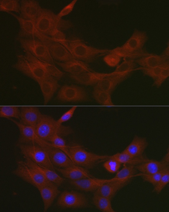
Immunofluorescence analysis of PC-12 cells using TRAF7 Rabbit pAb (CAB3095) at dilution of 1:50. Secondary antibody: Cy3-conjugated Goat anti-Rabbit IgG (H+L) (CABS007) at 1:500 dilution. Blue: DAPI for nuclear staining.