The UQCRC2 Monoclonal Antibody (CAB4366) is a high-quality antibody developed for reliable detection and analysis of target proteins. This antibody is produced using rabbit monoclonal technology, resulting in high specificity and sensitivity for detecting UQCRC2 in human samples. Validated for use in a variety of applications, including Western blot and immunohistochemistry, this antibody binds specifically to UQCRC2, allowing for precise detection and analysis in different cell types and tissues. Its reliability and consistency make it an ideal choice for studies in mitochondrial function, bioenergetics, and oxidative phosphorylation.
This antibody is validated for use in WB, IHC-P, ELISA applications and has demonstrated reactivity against Human, Mouse, Rat samples.
Product Name:
UQCRC2 Monoclonal Antibody
SKU:
CAB4366
Size:
20μL, 100μL
Reactivity:
Human, Mouse, Rat
Clone Number:
ARC0982
Conjugate:
Unconjugated
Immunogen:
Synthetic peptide. This information is considered to be commercially sensitive.
Recommended starting concentration is 1 μg/mL. Please optimize the concentration based on your specific assay requirements.
Synonyms:
QCR2, UQCR2, MC3DN5, UQCRC2
Positive Sample:
293T, Hep G2, A549, Mouse heart, Rat brain, Rat heart
Cellular Localization:
Mitochondrion Inner Membrane.
Calculated MW:
48kDa
Observed MW:
48kDa
The protein encoded by this gene is located in the mitochondrion, where it is part of the ubiquinol-cytochrome c reductase complex (also known as complex III). This complex constitutes a part of the mitochondrial respiratory chain. Defects in this gene are a cause of mitochondrial complex III deficiency nuclear type 5.
Purification Method
Affinity purification
Gene ID
7385
RRID
AB_2863250
Buffer Information
Store at -20℃. Avoid freeze / thaw cycles. Buffer: PBS containing 50% glycerol and 0.05% BSA, preserved with proclin300 or sodium azide, pH 7.3.
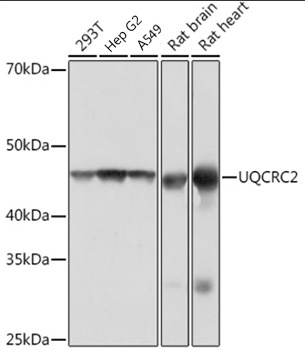
Western blot analysis of various lysates using UQCRC2 Rabbit mAb (CAB4366) at 1:1000 dilution. Secondary antibody: HRP-conjugated Goat anti-Rabbit IgG (H+L) (CABS014) at 1:10000 dilution. Lysates/proteins: 25μg per lane. Blocking buffer: 3% nonfat dry milk in TBST. Detection: ECL Basic Kit (AbGn00020). Exposure time: 1s.
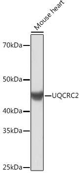
Western blot analysis of lysates from Mouse heart, using UQCRC2 Rabbit mAb (CAB4366) at 1:1000 dilution. Secondary antibody: HRP-conjugated Goat anti-Rabbit IgG (H+L) (CABS014) at 1:10000 dilution. Lysates/proteins: 25μg per lane. Blocking buffer: 3% nonfat dry milk in TBST. Detection: ECL Basic Kit (AbGn00020). Exposure time: 3min.
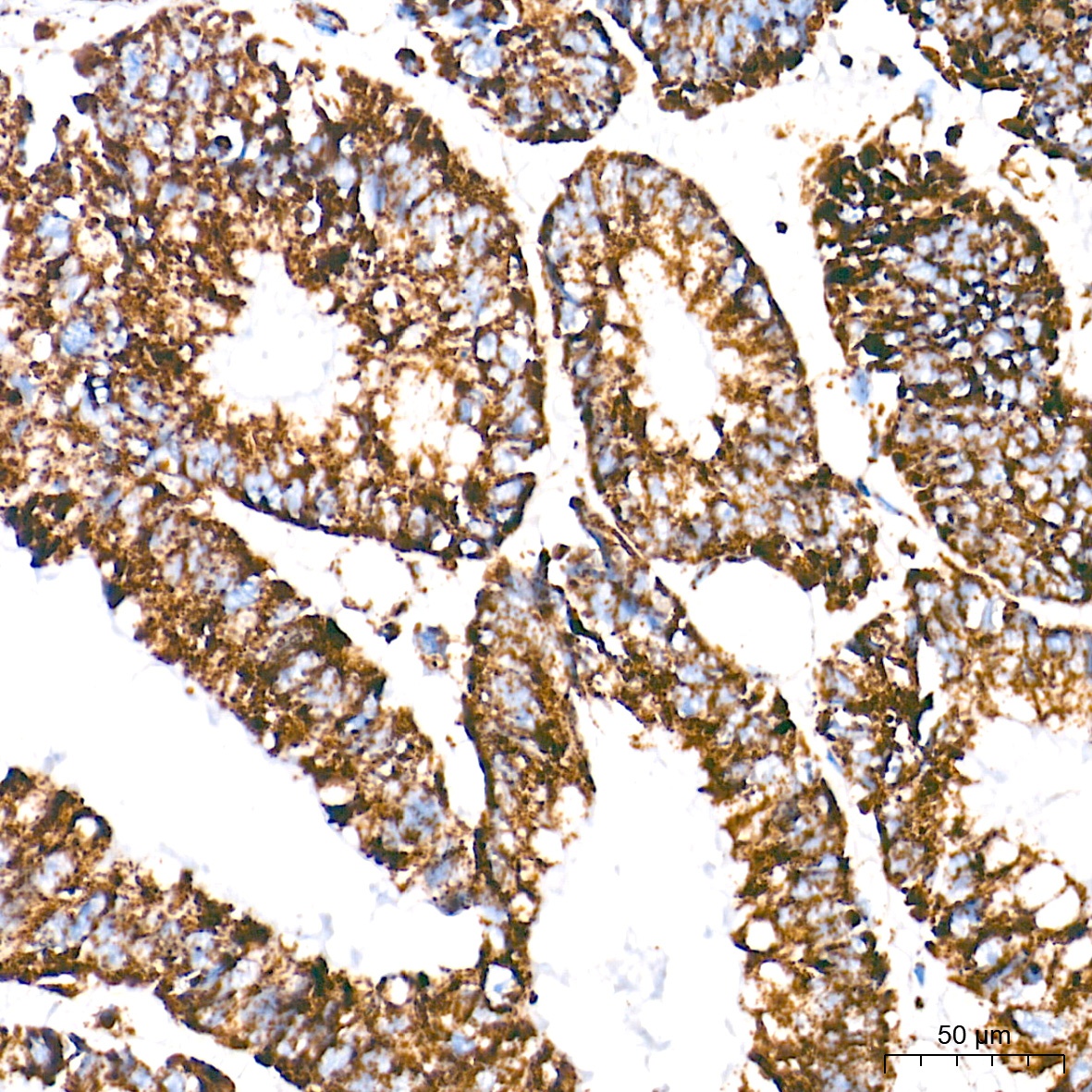
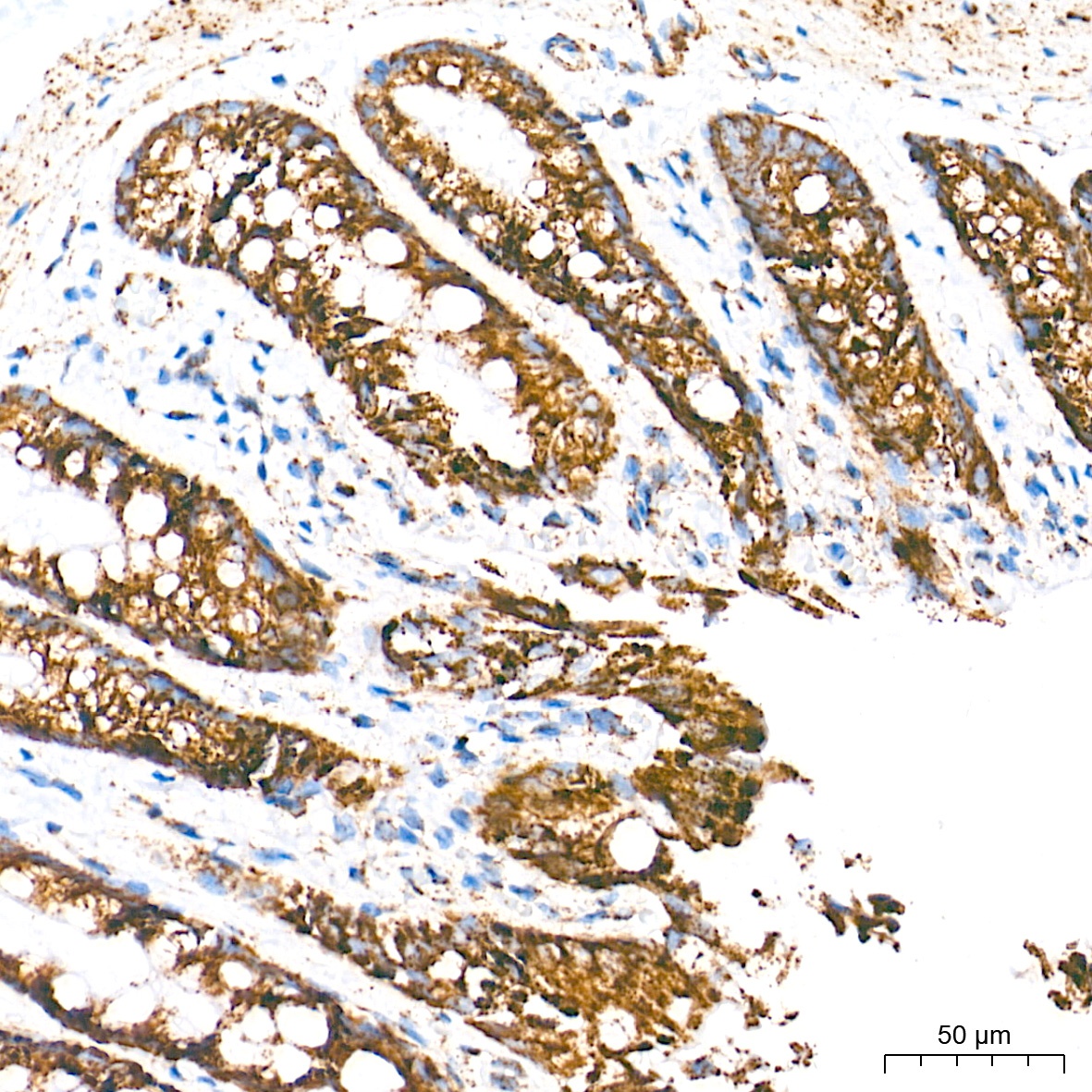
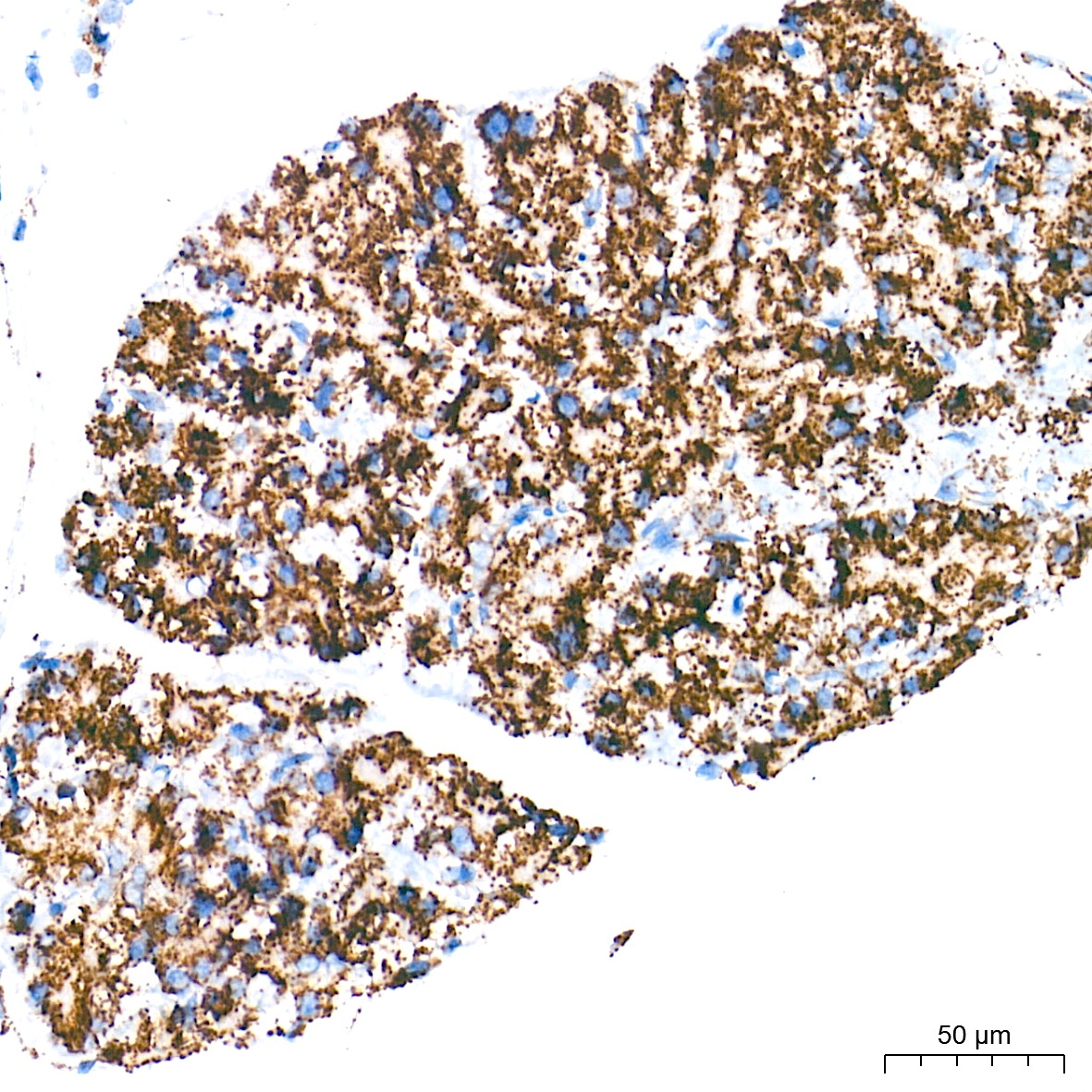
Immunohistochemistry analysis of paraffin-embedded Human colon carcinoma tissue using UQCRC2 Rabbit mAb (CAB4366) at a dilution of 1:500 (40x lens). High pressure antigen retrieval performed with 0.01M Tris-EDTA Buffer (pH 9.0) prior to IHC staining.
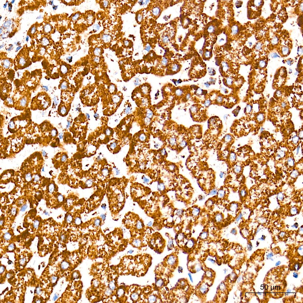
Immunohistochemistry analysis of paraffin-embedded Human liver tissue using UQCRC2 Rabbit mAb (CAB4366) at a dilution of 1:500 (40x lens). High pressure antigen retrieval performed with 0.01M Tris-EDTA Buffer (pH 9.0) prior to IHC staining.
Immunohistochemistry analysis of paraffin-embedded Rat colon tissue using UQCRC2 Rabbit mAb (CAB4366) at a dilution of 1:500 (40x lens). High pressure antigen retrieval performed with 0.01M Tris-EDTA Buffer (pH 9.0) prior to IHC staining.
Immunohistochemistry analysis of paraffin-embedded Rat pancreas tissue using UQCRC2 Rabbit mAb (CAB4366) at a dilution of 1:500 (40x lens). High pressure antigen retrieval performed with 0.01M Tris-EDTA Buffer (pH 9.0) prior to IHC staining.