The WWP1 Antibody (CAB5269) is a high-quality antibody developed for reliable detection and analysis of target proteins. This antibody, generated in rabbits, exhibits high specificity and reactivity with human samples, making it suitable for use in Western blot applications. By binding to the WWP1 protein, researchers can effectively study protein turnover and degradation processes in various cell types, providing valuable insights into cellular function and pathway regulation.WWP1, a HECT-type E3 ubiquitin ligase, plays a crucial role in targeting specific proteins for ubiquitination and subsequent degradation, thus influencing various cellular processes such as cell cycle regulation, signal transduction, and DNA repair.
This antibody is validated for use in WB, ELISA applications and has demonstrated reactivity against Human, Mouse, Rat samples.
Product Name:
WWP1 Antibody
SKU:
CAB5269
Size:
20μL, 100μL
Reactivity:
Human, Mouse, Rat
Conjugate:
Unconjugated
Immunogen:
Recombinant protein (or fragment).This information is considered to be commercially sensitive.
WW domain-containing proteins are found in all eukaryotes and play an important role in the regulation of a wide variety of cellular functions such as protein degradation, transcription, and RNA splicing. This gene encodes a protein which contains 4 tandem WW domains and a HECT (homologous to the E6-associated protein carboxyl terminus) domain. The encoded protein belongs to a family of NEDD4-like proteins, which are E3 ubiquitin-ligase molecules and regulate key trafficking decisions, including targeting of proteins to proteosomes or lysosomes. Alternative splicing of this gene generates at least 6 transcript variants; however, the full length nature of these transcripts has not been defined.
Purification Method
Affinity purification
Gene ID
11059
RRID
AB_2766090
Buffer Information
Store at -20℃. Avoid freeze / thaw cycles. Buffer: Buffer: PBS containing 50% glycerol, preserved with proclin300 or sodium azide, pH 7.3.
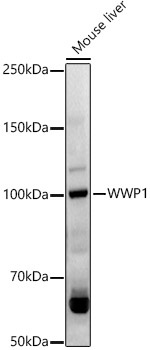
Western blot analysis of lysates from Mouse liver, using WWP1 Rabbit pAb (CAB5269) at 1:1000 dilution. Secondary antibody: HRP-conjugated Goat anti-Rabbit IgG (H+L) (CABS014) at 1:10000 dilution. Lysates/proteins: 25μg per lane. Blocking buffer: 3% nonfat dry milk in TBST. Detection: ECL Basic Kit (AbGn00020). Exposure time: 90s.
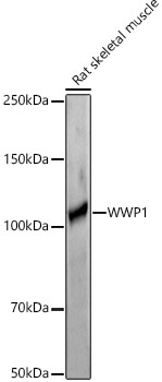
Western blot analysis of lysates from Rat skeletal muscle, using WWP1 Rabbit pAb (CAB5269) at 1:1000 dilution. Secondary antibody: HRP-conjugated Goat anti-Rabbit IgG (H+L) (CABS014) at 1:10000 dilution. Lysates/proteins: 25μg per lane. Blocking buffer: 3% nonfat dry milk in TBST. Detection: ECL Basic Kit (AbGn00020). Exposure time: 180s.