The AKR1C2 Antibody (CAB1048) is a high-quality antibody developed for reliable detection and analysis of target proteins. The antibody, produced in rabbits, exhibits high specificity and sensitivity when detecting AKR1C2 protein in human samples. Validated for use in Western blotting applications, it allows for accurate analysis of AKR1C2 expression in various cell types.AKR1C2, a member of the aldo-keto reductase family, plays a crucial role in the metabolism of endogenous and exogenous compounds, making it a key target for research in cancer biology, endocrinology, and drug metabolism studies.
This antibody is validated for use in WB, IHC-P, IF/ICC, ELISA applications and has demonstrated reactivity against Human, Mouse, Rat samples.
Product Name:
AKR1C2 Antibody
SKU:
CAB1048
Size:
20μL, 100μL
Reactivity:
Human, Mouse, Rat
Conjugate:
Unconjugated
Immunogen:
Recombinant protein (or fragment).This information is considered to be commercially sensitive.
This gene encodes a member of the aldo/keto reductase superfamily, which consists of more than 40 known enzymes and proteins. These enzymes catalyze the conversion of aldehydes and ketones to their corresponding alcohols using NADH and/or NADPH as cofactors. The enzymes display overlapping but distinct substrate specificity. This enzyme binds bile acid with high affinity, and shows minimal 3-alpha-hydroxysteroid dehydrogenase activity. This gene shares high sequence identity with three other gene members and is clustered with those three genes at chromosome 10p15-p14. Three transcript variants encoding two different isoforms have been found for this gene.
Purification Method
Affinity purification
Gene ID
1646
RRID
AB_2758028
Buffer Information
Store at -20℃. Avoid freeze / thaw cycles. Buffer: PBS containing 50% glycerol, preserved with proclin300 or sodium azide, pH 7.3.
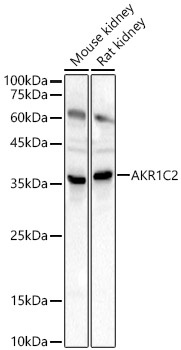
Western blot analysis of various lysates, using AKR1C2 Rabbit pAb (CAB1048) at 1:2000 dilution. Secondary antibody: HRP-conjugated Goat anti-Rabbit IgG (H+L) (CABS014) at 1:10000 dilution. Lysates/proteins: 25μg per lane. Blocking buffer: 3% nonfat dry milk in TBST. Detection: ECL Basic Kit (AbGn00020). Exposure time: 10s.
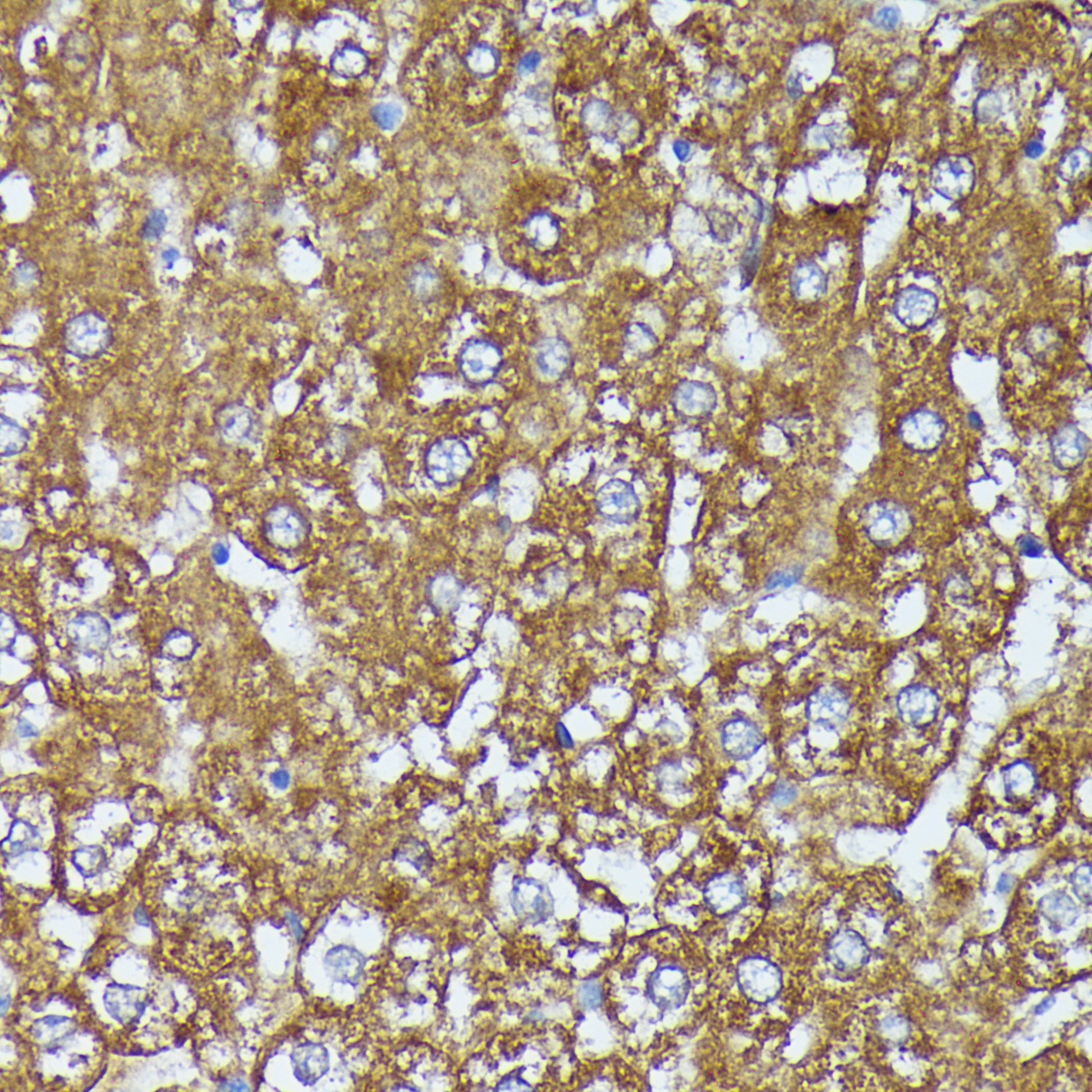
Immunohistochemistry analysis of paraffin-embedded Rat liver using AKR1C2 Rabbit pAb (CAB1048) at dilution of 1:100 (40x lens). Microwave antigen retrieval performed with 0.01M PBS Buffer (pH 7.2) prior to IHC staining.
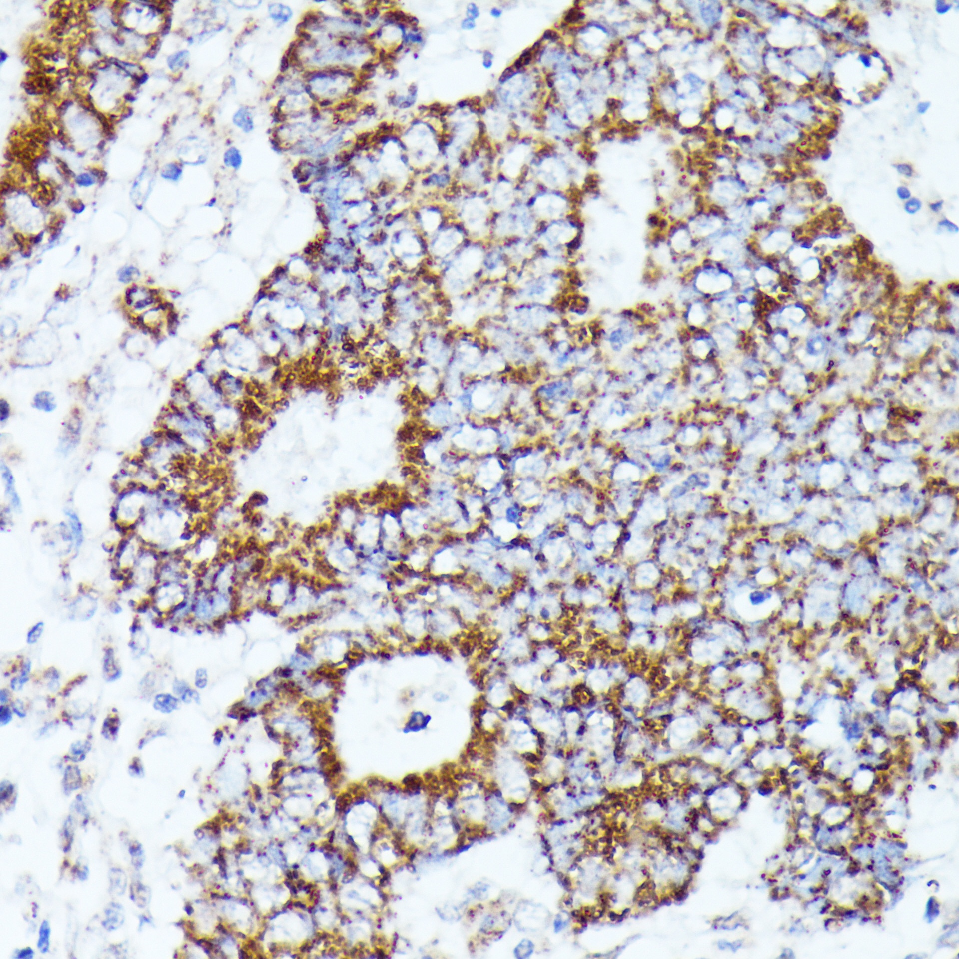
Immunohistochemistry analysis of paraffin-embedded Human Colon cancer using AKR1C2 Rabbit pAb (CAB1048) at dilution of 1:100 (40x lens). Microwave antigen retrieval performed with 0.01M PBS Buffer (pH 7.2) prior to IHC staining.
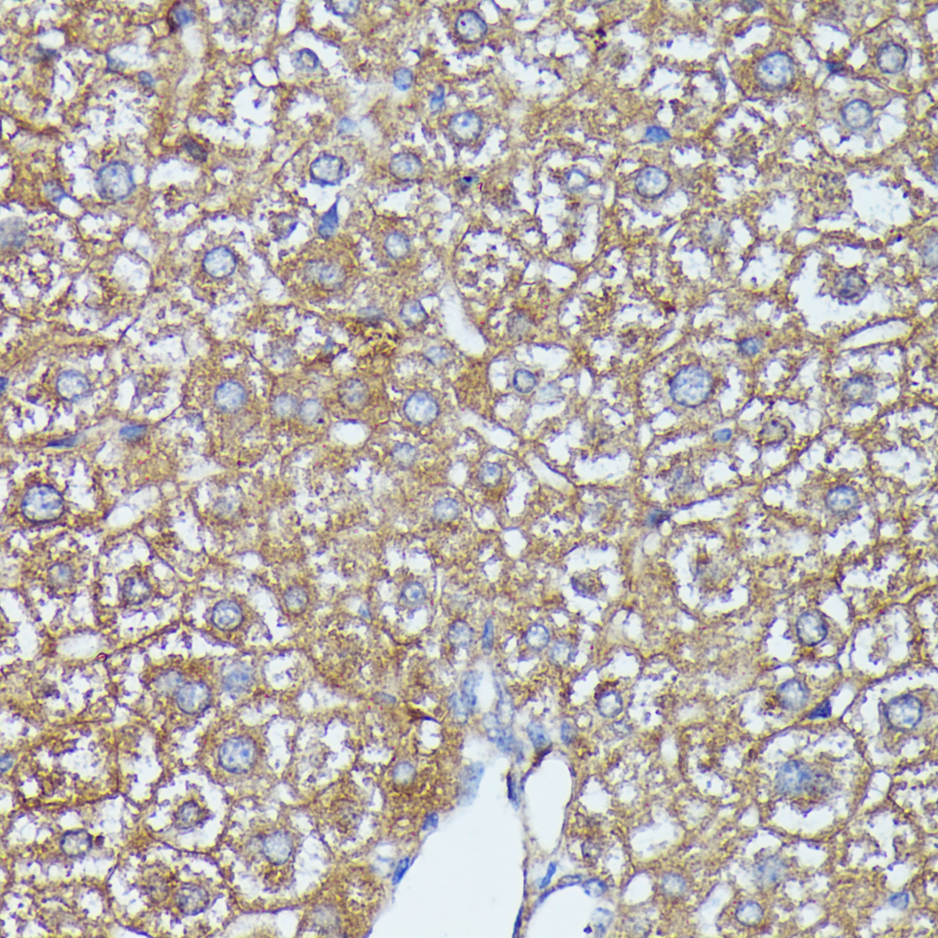
Immunohistochemistry analysis of paraffin-embedded Mouse liver using AKR1C2 Rabbit pAb (CAB1048) at dilution of 1:100 (40x lens). Microwave antigen retrieval performed with 0.01M PBS Buffer (pH 7.2) prior to IHC staining.
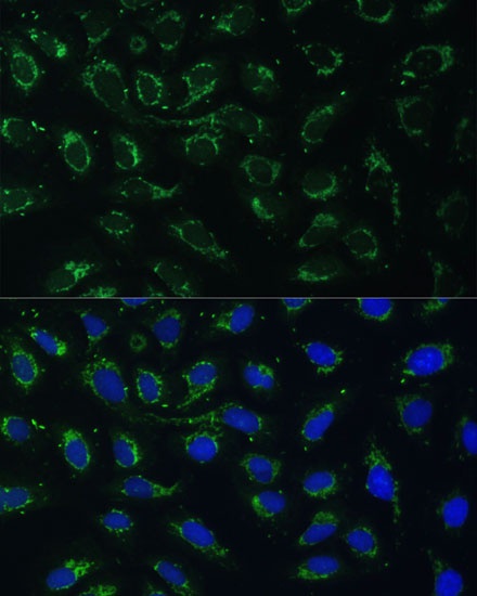
Immunofluorescence analysis of U-2 OS cells using AKR1C2 Rabbit pAb (CAB1048) at dilution of 1:100 (40x lens). Secondary antibody: Cy3-conjugated Goat anti-Rabbit IgG (H+L) (CABS007) at 1:500 dilution. Blue: DAPI for nuclear staining.