The Amphiregulin Antibody (CAB1860) is a high-quality antibody developed for reliable detection and analysis of target proteins. Amphiregulin is a member of the epidermal growth factor family and is known for its role in cell proliferation, differentiation, and migration.This polyclonal antibody, raised in rabbits, is highly specific and reactive with human samples. It is suitable for use in Western blot applications, allowing for the detection and analysis of amphiregulin protein in different cell types.
This antibody is validated for use in WB, ELISA applications and has demonstrated reactivity against Human, Mouse samples.
Product Name:
Amphiregulin Antibody
SKU:
CAB1860
Size:
20μL, 100μL
Reactivity:
Human, Mouse
Conjugate:
Unconjugated
Immunogen:
Recombinant protein (or fragment).This information is considered to be commercially sensitive.
Recommended starting concentration is 1 μg/mL. Please optimize the concentration based on your specific assay requirements.
Synonyms:
AR, SDGF, AREGB, CRDGF, Amphiregulin
Positive Sample:
A-549, Mouse brain
Cellular Localization:
Membrane, Single-Pass Membrane Protein.
Calculated MW:
28kDa
Observed MW:
43kDa
The protein encoded by this gene is a member of the epidermal growth factor family. It is an autocrine growth factor as well as a mitogen for astrocytes, Schwann cells and fibroblasts. It is related to epidermal growth factor (EGF) and transforming growth factor alpha (TGF-alpha). The protein interacts with the EGF/TGF-alpha receptor to promote the growth of normal epithelial cells, and it inhibits the growth of certain aggressive carcinoma cell lines. It also functions in mammary gland, oocyte and bone tissue development. This gene is associated with a psoriasis-like skin phenotype, and is also associated with other pathological disorders, including various types of cancers and inflammatory conditions.
Purification Method
Affinity purification
Gene ID
374
RRID
AB_2763895
Buffer Information
Store at -20℃. Avoid freeze / thaw cycles. Buffer: PBS with 0.01% thimerosal,50% glycerol,pH7.3.
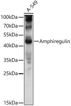
Western blot analysis of lysates from A-549 cells, using Amphiregulin Rabbit pAb (CAB1860) at 1:1000 dilution. Secondary antibody: HRP-conjugated Goat anti-Rabbit IgG (H+L) (CABS014) at 1:10000 dilution. Lysates/proteins: 25μg per lane. Blocking buffer: 3% nonfat dry milk in TBST. Detection: ECL Basic Kit (AbGn00020). Exposure time: 3s.
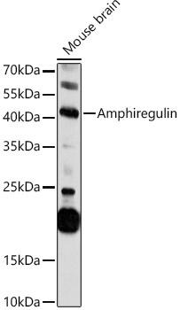
Western blot analysis of lysates from Mouse brain, using Amphiregulin Rabbit pAb (CAB1860) at 1:1000 dilution. Secondary antibody: HRP-conjugated Goat anti-Rabbit IgG (H+L) (CABS014) at 1:10000 dilution. Lysates/proteins: 25μg per lane. Blocking buffer: 3% nonfat dry milk in TBST. Detection: ECL Basic Kit (AbGn00020). Exposure time: 300s.