The NDUFB8 Antibody (CAB20457) is a high-quality antibody developed for reliable detection and analysis of target proteins. This antibody, produced in rabbits, is highly specific for human samples and is validated for use in Western blot applications. By binding to NDUFB8, the antibody enables precise detection and analysis of this important protein in various cell types.NDUFB8 is essential for the assembly and function of complex I in the electron transport chain, making it a key player in cellular energy production. Dysregulation of complex I has been implicated in various human diseases, including neurodegenerative disorders and metabolic syndromes.
This antibody is validated for use in WB, IHC-P, IF/ICC, ELISA applications and has demonstrated reactivity against Human, Mouse, Rat samples.
Product Name:
NDUFB8 Antibody
SKU:
CAB20457
Size:
20μL, 100μL
Reactivity:
Human, Mouse, Rat
Conjugate:
Unconjugated
Immunogen:
Recombinant protein (or fragment).This information is considered to be commercially sensitive.
Involved in mitochondrial respiratory chain complex I assembly. Located in endoplasmic reticulum and mitochondrion. Part of mitochondrial respiratory chain complex I. Implicated in nuclear type mitochondrial complex I deficiency 32. Biomarker of Alzheimer's disease and Parkinson's disease.
Purification Method
Affinity purification
Gene ID
4714
Buffer Information
Store at -20℃. Avoid freeze / thaw cycles. Buffer: PBS containing 50% glycerol, preserved with proclin300 or sodium azide, pH 7.3.
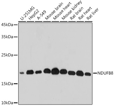
Western blot analysis of various lysates using NDUFB8 Rabbit pAb (CAB20457) at 1:500 dilution. Secondary antibody: HRP-conjugated Goat anti-Rabbit IgG (H+L) (CABS014) at 1:10000 dilution. Lysates/proteins: 25μg per lane. Blocking buffer: 3% nonfat dry milk in TBST. Detection: ECL Basic Kit (AbGn00020). Exposure time: 10s.
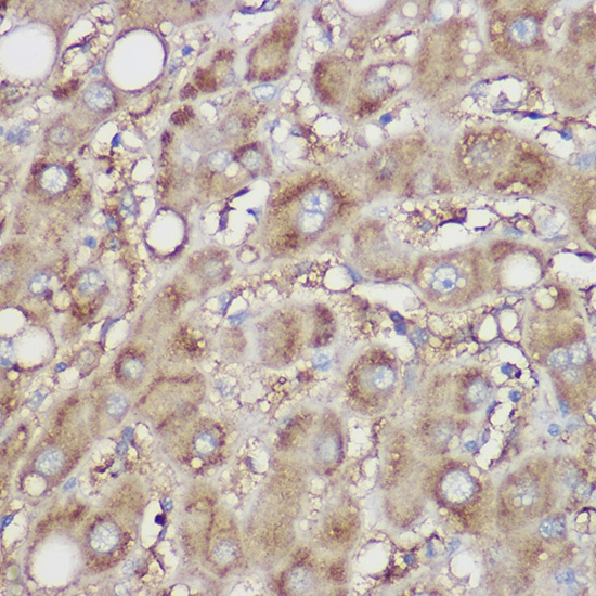
Immunohistochemistry analysis of paraffin-embedded Human liver using NDUFB8 Rabbit pAb (CAB20457) at dilution of 1:100 (40x lens). High pressure antigen retrieval performed with 0.01M Citrate buffer (pH 6.0) prior to IHC staining.
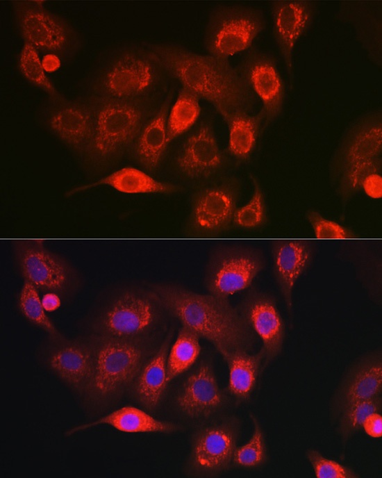
Immunofluorescence analysis of A-549 cells using NDUFB8 Rabbit pAb (CAB20457) at dilution of 1:100 (40x lens). Secondary antibody: Cy3-conjugated Goat anti-Rabbit IgG (H+L) (CABS007) at 1:500 dilution. Blue: DAPI for nuclear staining.
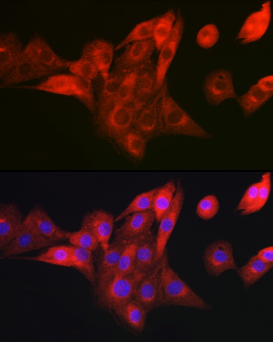
Immunofluorescence analysis of NIH/3T3 cells using NDUFB8 Rabbit pAb (CAB20457) at dilution of 1:100 (40x lens). Secondary antibody: Cy3-conjugated Goat anti-Rabbit IgG (H+L) (CABS007) at 1:500 dilution. Blue: DAPI for nuclear staining.