The NDUFS4 Monoclonal Antibody (CAB8691) is a high-quality antibody developed for reliable detection and analysis of target proteins. This polyclonal antibody, generated in rabbits, has high specificity for human samples and is optimized for use in Western blotting techniques. By targeting the NDUFS4 protein, this antibody enables researchers to investigate the role of mitochondrial complex I in various cell types, offering insights into bioenergetics and mitochondrial dysfunction in diseases such as neurodegenerative disorders and metabolic conditions.NDUFS4 is essential for maintaining mitochondrial function and energy production, making it a key player in cellular metabolism and ATP generation.
This antibody is validated for use in WB, IHC-P, IF/ICC, ELISA applications and has demonstrated reactivity against Human, Mouse, Rat samples.
Product Name:
NDUFS4 Monoclonal Antibody
SKU:
CAB8691
Size:
20μL, 100μL
Reactivity:
Human, Mouse, Rat
Clone Number:
ARC1784
Conjugate:
Unconjugated
Immunogen:
Recombinant protein (or fragment).This information is considered to be commercially sensitive.
This gene encodes an nuclear-encoded accessory subunit of the mitochondrial membrane respiratory chain NADH dehydrogenase (complex I, or NADH:ubiquinone oxidoreductase). Complex I removes electrons from NADH and passes them to the electron acceptor ubiquinone. Mutations in this gene can cause mitochondrial complex I deficiencies such as Leigh syndrome. Alternative splicing results in multiple transcript variants.
Purification Method
Affinity purification
Gene ID
4724
RRID
AB_2863585
Buffer Information
Store at -20℃. Avoid freeze / thaw cycles. Buffer: PBS containing 50% glycerol and 0.05% BSA, preserved with proclin300 or sodium azide, pH 7.3.
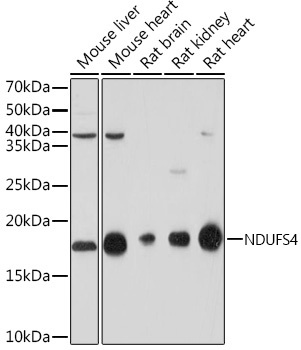
Western blot analysis of various lysates using NDUFS4 Rabbit mAb (CAB8691) at 1:1000 dilution. Secondary antibody: HRP-conjugated Goat anti-Rabbit IgG (H+L) (CABS014) at 1:10000 dilution. Lysates/proteins: 25μg per lane. Blocking buffer: 3% nonfat dry milk in TBST. Detection: ECL Basic Kit (AbGn00020). Exposure time: 30s.
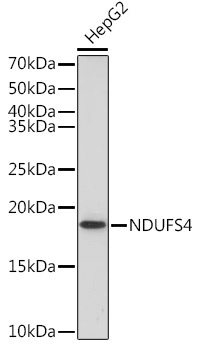
Western blot analysis of lysates from HepG2 cells, using NDUFS4 Rabbit mAb (CAB8691) at 1:1000 dilution. Secondary antibody: HRP-conjugated Goat anti-Rabbit IgG (H+L) (CABS014) at 1:10000 dilution. Lysates/proteins: 25μg per lane. Blocking buffer: 3% nonfat dry milk in TBST. Detection: ECL Basic Kit (AbGn00020). Exposure time: 90s.
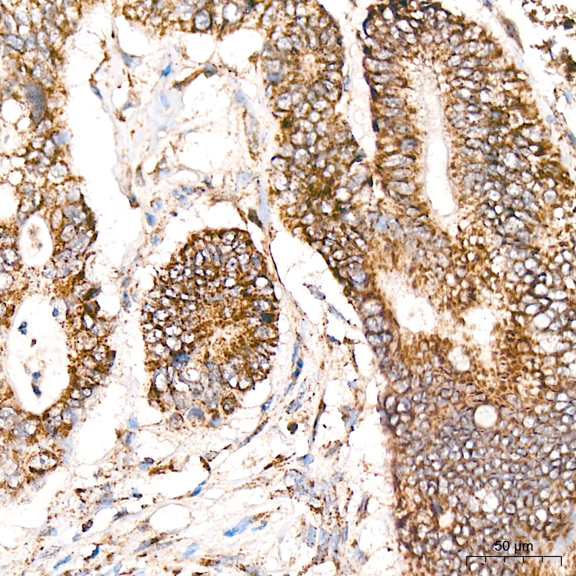
Immunohistochemistry analysis of paraffin-embedded Human colon carcinoma tissue using NDUFS4 Rabbit mAb (CAB8691) at a dilution of 1:400 (40x lens). High pressure antigen retrieval was performed with 0.01 M citrate buffer (pH 6.0) prior to IHC staining.
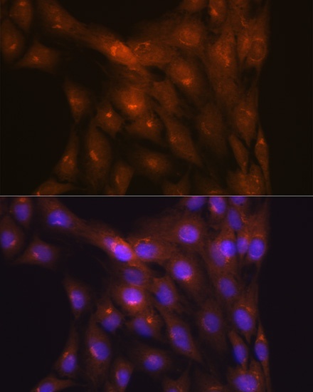
Immunofluorescence analysis of C6 cells using NDUFS4 Rabbit mAb (CAB8691) at dilution of 1:100 (40x lens). Secondary antibody: Cy3-conjugated Goat anti-Rabbit IgG (H+L) (CABS007) at 1:500 dilution. Blue: DAPI for nuclear staining.


![Anti-NDUFS4 [R08-4O5] Monoclonal Antibody (AGMB03108)](https://cdn11.bigcommerce.com/s-h68l9z2lnx/images/stencil/590x590/products/274397/678277/anti-ndufs4-r08-4o5-monoclonal-antibody-agmb03108__22940.1773035291.jpg?c=2 "Anti-NDUFS4 [R08-4O5] Monoclonal Antibody (AGMB03108)")

