The Phospho-RIPK1/RIP-S166 Antibody (CABP1230) is a high-quality antibody developed for reliable detection and analysis of target proteins. This antibody, developed and validated for use in Western blot applications, specifically detects the phosphorylated form of RIPK1 at serine 166. RIPK1 is a crucial regulator of cell survival and death, and dysregulation of its activity has been implicated in various diseases, including cancer, neurodegenerative disorders, and inflammatory conditions. By targeting the phosphorylated form of RIPK1, researchers can gain insights into the intricate signaling pathways that govern cell fate decisions in these disease contexts.
This antibody is validated for use in WB, ELISA applications and has demonstrated reactivity against Human, Mouse samples.
Product Name:
Phospho-RIPK1/RIP-S166 Antibody
SKU:
CABP1230
Size:
20μL, 100μL
Reactivity:
Human, Mouse
Conjugate:
Unconjugated
Immunogen:
Synthetic peptide. This information is considered to be commercially sensitive.
Sequence:
MWSK L
Tested Applications:
WBELISA
Recommended Dilution:
WB
1:500 - 1:5000
ELISA
Recommended starting concentration is 1 μg/mL. Please optimize the concentration based on your specific assay requirements.
This gene encodes a member of the receptor-interacting protein (RIP) family of serine/threonine protein kinases. The encoded protein plays a role in inflammation and cell death in response to tissue damage, pathogen recognition, and as part of developmental regulation. RIPK1/RIPK3 kinase-mediated necrosis is referred to as necroptosis. Genetic disruption of this gene in mice results in death shortly after birth.
Purification Method
Affinity purification
Gene ID
8737
Buffer Information
Store at -20℃. Avoid freeze / thaw cycles. Buffer: PBS with 0.09% Sodium azide,50% glycerol,pH7.3.
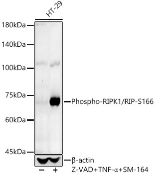
Western blot analysis of lysates from HT-29 cells, using Phospho-RIPK1/RIP-S166 Rabbit pAb (CABP1230) at 1:800 dilution. HT-29 cells were treated with Z-VAD (50uM), human TNF-α (50ng/ml) and SM-164 (200nM) at 37℃ for 5 hour. Secondary antibody: HRP-conjugated Goat anti-Rabbit IgG (H+L) (CABS014) at 1:10000 dilution. Lysates/proteins: 25μg per lane. Blocking buffer: 3% nonfat dry milk in TBST. Detection: ECL Basic Kit (AbGn00020). Exposure time: 60s.