The AQP8 Antibody (CAB8539) is a high-quality antibody developed for reliable detection and analysis of target proteins. This antibody, produced in rabbits, is highly specific for Aquaporin 8 in human samples and is validated for use in various applications, including Western blotting.Aquaporin 8 is known for its role in the transport of water across cell membranes, particularly in the gastrointestinal tract and kidney. Its expression is crucial for maintaining water balance in the body and for proper functioning of these organs.
This antibody is validated for use in WB, IF/ICC, ELISA applications and has demonstrated reactivity against Human, Mouse, Rat samples.
Product Name:
AQP8 Antibody
SKU:
CAB8539
Size:
20μL, 100μL
Reactivity:
Human, Mouse, Rat
Conjugate:
Unconjugated
Immunogen:
Synthetic peptide. This information is considered to be commercially sensitive.
Recommended starting concentration is 1 μg/mL. Please optimize the concentration based on your specific assay requirements.
Synonyms:
AQP-8, AQP8
Positive Sample:
Human liver, Human heart, Rat liver
Cellular Localization:
Membrane, Multi-Pass Membrane Protein.
Calculated MW:
27kDa
Observed MW:
27kDa
Aquaporin 8 (AQP8) is a water channel protein. Aquaporins are a family of small integral membrane proteins related to the major intrinsic protein (MIP or AQP0). Aquaporin 8 mRNA is found in pancreas and colon but not other tissues.
Purification Method
Affinity purification
Gene ID
343
RRID
AB_2768409
Buffer Information
Store at -20℃. Avoid freeze / thaw cycles. Buffer: PBS containing 50% glycerol, preserved with proclin300 or sodium azide, pH 7.3.
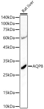
Western blot analysis of lysates from Rat liver, using AQP8 Rabbit pAb (CAB8539) at 1:3000 dilution. Secondary antibody: HRP-conjugated Goat anti-Rabbit IgG (H+L) (CABS014) at 1:10000 dilution. Lysates/proteins: 25μg per lane. Blocking buffer: 3% nonfat dry milk in TBST. Detection: ECL Basic Kit (AbGn00020). Exposure time: 30s.
Western blot analysis of various lysates using AQP8 Rabbit pAb (CAB8539) at 1:1000 dilution. Secondary antibody: HRP-conjugated Goat anti-Rabbit IgG (H+L) (CABS014) at 1:10000 dilution. Lysates / proteins: 25 μg per lane. Blocking buffer: 3 % nonfat dry milk in TBST. Detection: ECL Basic Kit (AbGn00020). Exposure time: 90s.
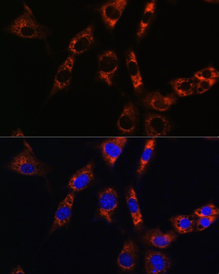
Immunofluorescence analysis of NIH/3T3 cells using AQP8 Rabbit pAb (CAB8539) at dilution of 1:100. Secondary antibody: Cy3-conjugated Goat anti-Rabbit IgG (H+L) (CABS007) at 1:500 dilution. Blue: DAPI for nuclear staining.