
Guide to Fluorescence Activated Cell Sorting (FACs): The Latest Technology in Cell Separation
Guide to Fluorescence-Activated Cell Sorting (FACS)
Fluorescence-activated cell sorting (FACS) is a specialised form of flow cytometry that physically isolates individual cells from a mixed sample based on their fluorescence and light-scattering properties. With sorting purities approaching 99.99%, it is one of the most powerful tools in modern cell biology, immunology and stem cell research.
Explore flow cytometry antibodies →Key Takeaways
- FACS isolates individual cells with a purity approaching 99.99%.
- It sorts cells by size, granularity, surface markers and fluorescence intensity.
- Cells are labelled with fluorochrome-conjugated antibodies such as FITC, PE and APC.
- Charged droplets containing single cells are deflected into collection tubes by electric plates.
- Applications span cancer research, immunology, stem cell biology and clinical diagnostics.
- FACS is a sorting-capable subset of flow cytometry — all FACS is flow cytometry, but not all flow cytometry sorts.
Reagents for FACS & Flow Cytometry
From fluorochrome-conjugated marker antibodies to viability dyes and fixation buffers, these validated reagents support every stage of a cell-sorting experiment.
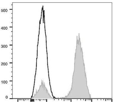
CD3 Monoclonal Antibody (FITC)
A pan-T-cell marker in a green fluorochrome for the first sorting channel.
View antibody →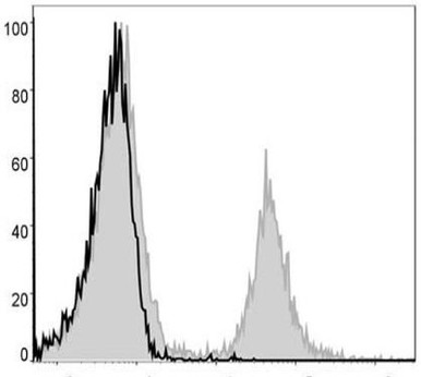
CD4 Monoclonal Antibody (PE-Cy5)
Identifies helper T cells for multi-colour sorting panels.
View antibody →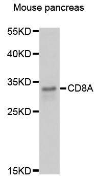
CD8 Monoclonal Antibody (APC)
A far-red conjugate for resolving cytotoxic T cells alongside CD3 and CD4.
View antibody →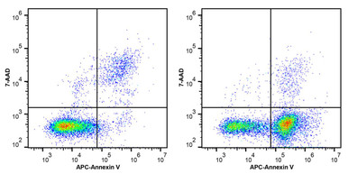
7-AAD Viability Staining Solution
Excludes dead cells so only viable events are sorted.
View product →
Intracellular Fix/Perm Buffer Kit
Fixes and permeabilises cells to stain intracellular targets.
View product →
Annexin V-FITC/PI Apoptosis Kit
Distinguishes live, apoptotic and necrotic cells during sorting.
View antibody →What is FACS?
Fluorescence-activated cell sorting is a technique for isolating individual cells from a heterogeneous sample and analysing their properties one cell at a time. It is a specialised, sorting-capable form of flow cytometry, and it is revolutionising cell biology by allowing researchers to purify defined cell populations with an accuracy of up to 99.99% — making it ideal for any application where cell purity matters.
The power of FACS lies in combining measurement and physical separation. As each cell passes a laser, the instrument records its size, internal complexity and the fluorescence of any bound markers, then diverts that individual cell into the correct collection tube — all in a fraction of a second.
How Does FACS Work?
A FACS instrument works by carrying cells single-file in a fast-moving fluid stream past one or more laser beams. As each cell crosses a laser, forward-scatter reports its size and side-scatter its internal granularity, while detectors capture the light emitted by fluorochrome-labelled markers on or inside the cell.
The stream is then broken into thousands of tiny droplets, each ideally containing a single cell. Droplets carrying a cell of interest are given an electric charge, and as they fall between two deflection plates, positively charged droplets are pulled toward the negative plate and negatively charged droplets toward the positive plate. The sorted cells are collected in separate tubes, while unwanted droplets pass straight into waste.
The FACS Protocol
FACS is a complex procedure requiring trained personnel and specialised equipment, but the core workflow is consistent. First, the cells are labelled with fluorescent dyes — usually antibodies conjugated to fluorochromes such as FITC (a green dye excited by blue light) or PE (a red dye excited by green light) that bind specific cell-surface markers.
The cells are incubated with the antibodies for 30 to 60 minutes, then washed and resuspended in a sorting buffer containing salts and proteins that keep the cells healthy during the run. A viability dye such as 7-AAD is often added to exclude dead cells. The prepared sample is then loaded onto the sorter, gated on the populations of interest, and sorted into collection tubes for downstream culture or analysis.
FACS vs Flow Cytometry
The two terms are often used interchangeably, but there is a key distinction. Flow cytometry is the broad technique of measuring the properties of cells in a fluid stream; it analyses cells but does not necessarily separate them. FACS adds the sorting capability — it not only measures each cell but also physically recovers defined populations for further use. In short, all FACS is flow cytometry, but only sorting-capable instruments perform FACS.
Applications of FACS
The ability to purify defined cell populations makes FACS indispensable across the life sciences.
Cancer research
FACS isolates tumour and immune cell subsets from complex tissues, enabling the study of cancer heterogeneity and the immune response to tumours.
Immunology
Researchers routinely sort T-cell, B-cell and other leukocyte subsets — using markers such as CD3, CD4 and CD8 — to dissect immune function.
Stem cell research
FACS purifies rare stem and progenitor populations from marrow or tissue based on their unique marker profiles.
Developmental biology
Sorting cells by lineage marker at successive stages reveals how cell populations arise and differentiate during development.
Build Your Sorting Panel with Assay Genie
Fluorochrome-conjugated CD antibodies, viability dyes, fixation buffers and apoptosis kits — everything needed to design a multi-colour FACS experiment, validated for flow.
Browse flow cytometry antibodies →Frequently Asked Questions
What does FACS stand for?
FACS stands for fluorescence-activated cell sorting, a technique that sorts individual cells based on their fluorescence and light-scattering properties.
How accurate is FACS sorting?
Modern FACS can achieve sort purities approaching 99.99%, making it ideal for applications requiring highly pure cell populations.
What is the difference between FACS and flow cytometry?
Flow cytometry measures cell properties in a stream; FACS additionally sorts and physically recovers defined populations. All FACS is flow cytometry, but not all flow cytometry sorts.
What reagents are needed for FACS?
Typically fluorochrome-conjugated antibodies against surface markers, a viability dye to exclude dead cells, and a sorting buffer; intracellular staining also needs a fixation and permeabilisation kit.
How long does antibody labelling take?
Cells are usually incubated with fluorochrome-conjugated antibodies for 30 to 60 minutes, then washed before sorting.
Seán is co-founder of Assay Genie and holds a PhD in cell biology, with a research background in flow cytometry and apoptosis.
Recent Posts
-
Suspected Plague Death at a Siberian Anti-Plague Institute: What Researchers Should Know
Infectious Disease · News AnalysisSuspected Plague Death at a Siberian Anti-Plague Institute: What …6th Oct 2026 -
LPS ELISA Troubleshooting: The Importance of Sample Nature
Quick answer A researcher measuring circulating lipopolysaccharide (LPS) in human serum …25th Sep 2026 -

Sekundärantikörper wählen: Die Regel, die im Labor wirklich zählt
Sekundärantikörper wählen: Die Regel, die im Labor wirklich zählt Wählen Sie einen Sekundärantikörp …24th Sep 2026