The CD68 Antibody (CAB13286) is a high-quality antibody developed for reliable detection and analysis of target proteins. This antibody, developed in rabbits, exhibits high reactivity with human samples and has been validated for use in Western blot applications. By binding specifically to the CD68 protein, it enables accurate detection and analysis in various cell types, making it ideal for investigations in immunology and cancer research.CD68 plays a crucial role in immune response and inflammation regulation, making it a key target for studies on diseases such as cancer, autoimmune disorders, and inflammatory conditions.
This antibody is validated for use in WB, IHC-P, IF/ICC, ELISA applications and has demonstrated reactivity against Human, Mouse samples.
Product Name:
CD68 Antibody
SKU:
CAB13286
Size:
20μL, 100μL
Reactivity:
Human, Mouse
Conjugate:
Unconjugated
Immunogen:
Synthetic peptide. This information is considered to be commercially sensitive.
Recommended starting concentration is 1 μg/mL. Please optimize the concentration based on your specific assay requirements.
Synonyms:
GP110, LAMP4, SCARD1, CD68
Positive Sample:
THP-1
Cellular Localization:
Cell Membrane, Endosome Membrane, Lysosome Membrane, Single-Pass Type I Membrane Protein, Single-Pass Type I Membrane Protein.
Calculated MW:
37kDa
Observed MW:
70-80kDa
This gene encodes a 110-kD transmembrane glycoprotein that is highly expressed by human monocytes and tissue macrophages. It is a member of the lysosomal/endosomal-associated membrane glycoprotein (LAMP) family. The protein primarily localizes to lysosomes and endosomes with a smaller fraction circulating to the cell surface. It is a type I integral membrane protein with a heavily glycosylated extracellular domain and binds to tissue- and organ-specific lectins or selectins. The protein is also a member of the scavenger receptor family. Scavenger receptors typically function to clear cellular debris, promote phagocytosis, and mediate the recruitment and activation of macrophages. Alternative splicing results in multiple transcripts encoding different isoforms.
Purification Method
Affinity purification
Gene ID
968
RRID
AB_2760143
Buffer Information
Store at -20℃. Avoid freeze / thaw cycles. Buffer: PBS containing 50% glycerol, preserved with proclin300 or sodium azide, pH 7.3.
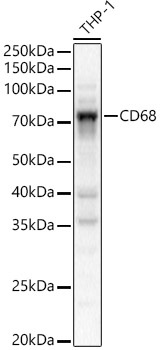
Western blot analysis of lysates from THP-1 cells, using CD68 Rabbit pAb (CAB13286) at 1:2000 dilution. Secondary antibody: HRP-conjugated Goat anti-Rabbit IgG (H+L) (CABS014) at 1:10000 dilution. Lysates/proteins: 25μg per lane. Blocking buffer: 3% nonfat dry milk in TBST. Detection: ECL Basic Kit (AbGn00020). Exposure time: 10s.
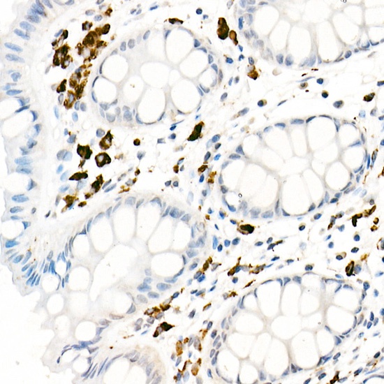
Immunohistochemistry analysis of paraffin-embedded Human colon using CD68 Rabbit pAb (CAB13286) at dilution of 1:20 (40x lens). High pressure antigen retrieval performed with 0.01M Citrate buffer (pH 6.0) prior to IHC staining.
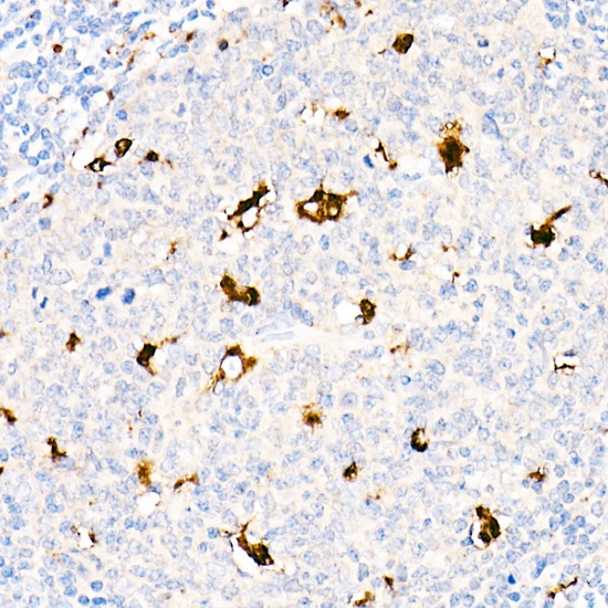
Immunohistochemistry analysis of paraffin-embedded Human tonsil using CD68 Rabbit pAb (CAB13286) at dilution of 1:20 (40x lens). High pressure antigen retrieval performed with 0.01M Citrate buffer (pH 6.0) prior to IHC staining.
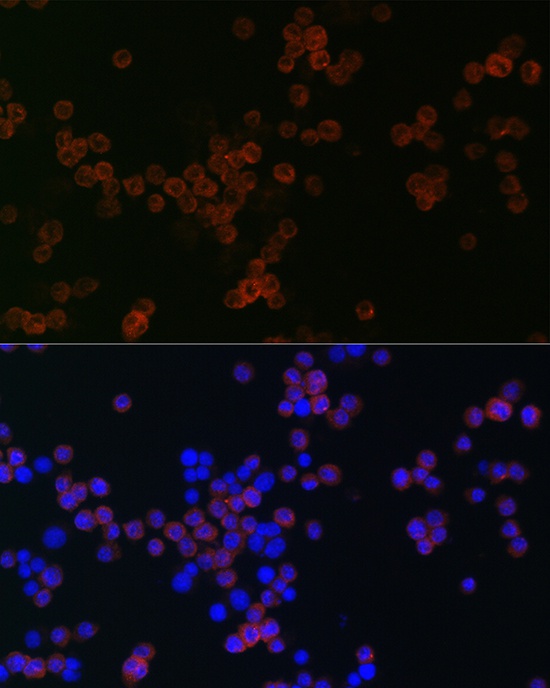
Immunofluorescence analysis of RAW264.7 cells using CD68 Rabbit pAb (CAB13286) at dilution of 1:50 (40x lens). Secondary antibody: Cy3-conjugated Goat anti-Rabbit IgG (H+L) (CABS007) at 1:500 dilution. Blue: DAPI for nuclear staining.


![Anti-CD68 [R04-7E1] Monoclonal Antibody (AGMB01535)](https://cdn11.bigcommerce.com/s-h68l9z2lnx/images/stencil/590x590/products/272824/677288/anti-cd68-r04-7e1-monoclonal-antibody-agmb01535__62388.1773032174.jpg?c=2 "Anti-CD68 [R04-7E1] Monoclonal Antibody (AGMB01535)")
![Anti-CD68 [R07-6G-9] Monoclonal Antibody (AGMB03783)](https://cdn11.bigcommerce.com/s-h68l9z2lnx/images/stencil/590x590/products/275072/676605/anti-cd68-r07-6g-9-monoclonal-antibody-agmb03783__38778.1773030010.jpg?c=2 "Anti-CD68 [R07-6G-9] Monoclonal Antibody (AGMB03783)")
![Anti-CD68 [1B8-8F6-6H8] Monoclonal Antibody (AGMB06310)](https://cdn11.bigcommerce.com/s-h68l9z2lnx/images/stencil/590x590/products/277591/680874/anti-cd68-1b8-8f6-6h8-monoclonal-antibody-agmb06310__95643.1773043461.jpg?c=2 "Anti-CD68 [1B8-8F6-6H8] Monoclonal Antibody (AGMB06310)")
![Anti-CD68 [R02-5K-4] Monoclonal Antibody (AGMB06514)](https://cdn11.bigcommerce.com/s-h68l9z2lnx/images/stencil/590x590/products/277795/733698/anti-cd68-r02-5k-4-monoclonal-antibody-agmb06514__86866.1777190843.jpg?c=2 "Anti-CD68 [R02-5K-4] Monoclonal Antibody (AGMB06514)")