The ERCC4 Antibody (CAB8119) is a high-quality antibody developed for reliable detection and analysis of target proteins. This antibody, produced in rabbits, exhibits high reactivity with human samples and has been validated for use in Western blot applications. By binding specifically to the ERCC4 protein, this antibody allows for the precise detection and analysis of ERCC4 in various cell types.ERCC4, also known as XPF, plays a crucial role in maintaining genomic stability by participating in the repair of DNA damage. Dysregulation of ERCC4 has been linked to various diseases, including cancer and neurodegenerative disorders.
This antibody is validated for use in WB, IP, ELISA applications and has demonstrated reactivity against Human, Mouse, Rat samples.
Product Name:
ERCC4 Antibody
SKU:
CAB8119
Size:
20μL, 100μL
Reactivity:
Human, Mouse, Rat
Conjugate:
Unconjugated
Immunogen:
Synthetic peptide. This information is considered to be commercially sensitive.
0.5μg-4μg antibody for 200μg-400μg extracts of whole cells
ELISA
Recommended starting concentration is 1 μg/mL. Please optimize the concentration based on your specific assay requirements.
Synonyms:
XPF, RAD1, FANCQ, XFEPS, ERCC11, ERCC4
Positive Sample:
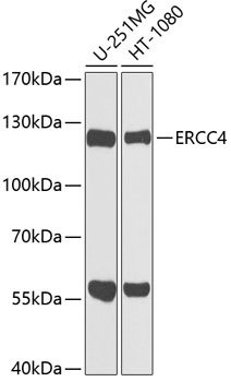
U-251MG, HT-1080
Cellular Localization:
Nucleus.
Calculated MW:
104kDa
Observed MW:
120kDa
The protein encoded by this gene forms a complex with ERCC1 and is involved in the 5' incision made during nucleotide excision repair. This complex is a structure specific DNA repair endonuclease that interacts with EME1. Defects in this gene are a cause of xeroderma pigmentosum complementation group F (XP-F), or xeroderma pigmentosum VI (XP6).
Purification Method
Affinity purification
Gene ID
2072
RRID
AB_2769352
Buffer Information
Store at -20℃. Avoid freeze / thaw cycles. Buffer: PBS containing 50% glycerol, preserved with proclin300 or sodium azide, pH 7.3.
Western blot analysis of various lysates using ERCC4 Rabbit pAb (CAB8119) at 1:1000 dilution. Secondary antibody: HRP-conjugated Goat anti-Rabbit IgG (H+L) (CABS014) at 1:10000 dilution. Lysates/proteins: 25μg per lane. Blocking buffer: 3% nonfat dry milk in TBST. Detection: ECL Basic Kit (AbGn00020). Exposure time: 30s.
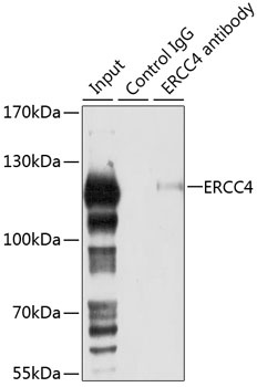
Immunoprecipitation analysis of 150 μg extracts of 293T cells using 3 μg ERCC4 antibody (CAB8119). Western blot was performed from the immunoprecipitate using ERCC4 antibody (CAB8119) at a dilution of 1:1000.