The ERVW-1 Antibody (CAB16522) is a high-quality antibody developed for reliable detection and analysis of target proteins. This antibody, produced in rabbits, is highly reactive with human samples and is validated for use in applications such as Western blotting. By binding to the ERVW-1 protein, this antibody allows for the detection and analysis of ERVW-1 in various types of cells, making it well-suited for studies in reproductive biology and infertility research.ERVW-1, also known as syncytin-1, plays a crucial role in the fusion of placental cells and is essential for the development of a healthy pregnancy.
This antibody is validated for use in WB, ELISA applications and has demonstrated reactivity against Human samples.
Product Name:
ERVW-1 Antibody
SKU:
CAB16522
Size:
20μL, 100μL
Reactivity:
Human
Conjugate:
Unconjugated
Immunogen:
Synthetic peptide. This information is considered to be commercially sensitive.
Cell Membrane, Peripheral Membrane Protein, Single-Pass Type I Membrane Protein, Virion.
Calculated MW:
60kDa
Observed MW:
60kDa
Many different human endogenous retrovirus (HERV) families are expressed in normal placental tissue at high levels, suggesting that HERVs are functionally important in reproduction. This gene is part of an HERV provirus on chromosome 7 that has inactivating mutations in the gag and pol genes. This gene is the envelope glycoprotein gene which appears to have been selectively preserved. The gene's protein product is expressed in the placental syncytiotrophoblast and is involved in fusion of the cytotrophoblast cells to form the syncytial layer of the placenta. The protein has the characteristics of a typical retroviral envelope protein, including a furin cleavage site that separates the surface (SU) and transmembrane (TM) proteins which form a heterodimer. Alternatively spliced transcript variants encoding the same protein have been found for this gene.
Purification Method
Affinity purification
Gene ID
30816
RRID
AB_2769360
Buffer Information
Store at -20℃. Avoid freeze / thaw cycles. Buffer: PBS containing 50% glycerol, preserved with proclin300 or sodium azide, pH 7.3.
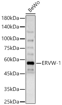
Western blot analysis of lysates from BeWo cells, using ERVW-1 Rabbit pAb (CAB16522) at 1:600 dilution. Secondary antibody: HRP-conjugated Goat anti-Rabbit IgG (H+L) (CABS014) at 1:10000 dilution. Lysates/proteins: 25μg per lane. Blocking buffer: 3% nonfat dry milk in TBST. Detection: ECL Basic Kit (AbGn00020). Exposure time: 90s.


 Antibody (HDBS0112)")
![Anti-Syntenin 1 [ZN69] Monoclonal Antibody (AGMB06263)](https://cdn11.bigcommerce.com/s-h68l9z2lnx/images/stencil/590x590/products/277544/677404/anti-syntenin-1-zn69-monoclonal-antibody-agmb06263__52812.1773032539.jpg?c=2 "Anti-Syntenin 1 [ZN69] Monoclonal Antibody (AGMB06263)")


 ELISA Kit (AEFI00327)")
 ELISA Kit (AEFI00327)")
![Purified Anti-Human CD95 Antibody [APO-1-1] (AGEL5073)](https://cdn11.bigcommerce.com/s-h68l9z2lnx/images/stencil/590x590/products/271077/690778/purified-anti-human-cd95-antibody-apo-1-1-agel5073__59712.1774500922.jpg?c=2 "Purified Anti-Human CD95 Antibody [APO-1-1] (AGEL5073)")