Glycophorin C (GYPC) Monoclonal Antibody (CAB11472)
The Glycophorin C (GYPC) Monoclonal Antibody (CAB11472) is a high-quality antibody developed for reliable detection and analysis of target proteins. This highly specific antibody, raised in rabbits, is suitable for use in various applications, including Western blot and immunohistochemistry.Glycophorin C is known for its role in maintaining the shape and flexibility of red blood cells, as well as its involvement in certain blood disorders. By targeting this protein, researchers can better understand its function and significance in health and disease.
This antibody is validated for use in WB, IHC-P, ELISA applications and has demonstrated reactivity against Human, Mouse, Rat samples.
Product Name:
Glycophorin C (GYPC) Monoclonal Antibody
SKU:
CAB11472
Size:
20μL, 100μL
Reactivity:
Human, Mouse, Rat
Clone Number:
ARC0605
Conjugate:
Unconjugated
Immunogen:
Synthetic peptide. This information is considered to be commercially sensitive.
Cell Membrane, Single-Pass Type Iii Membrane Protein.
Calculated MW:
14kDa
Observed MW:
30-40kDa
Glycophorin C (GYPC) is an integral membrane glycoprotein. It is a minor species carried by human erythrocytes, but plays an important role in regulating the mechanical stability of red cells. A number of glycophorin C mutations have been described. The Gerbich and Yus phenotypes are due to deletion of exon 3 and 2, respectively. The Webb and Duch antigens, also known as glycophorin D, result from single point mutations of the glycophorin C gene. The glycophorin C protein has very little homology with glycophorins A and B. Alternate splicing results in multiple transcript variants.
Purification Method
Affinity purification
Gene ID
2995
RRID
AB_2861573
Buffer Information
Store at -20℃. Avoid freeze / thaw cycles. Buffer: PBS containing 50% glycerol and 0.05% BSA, preserved with proclin300 or sodium azide, pH 7.3.
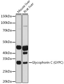
Western blot analysis of various lysates using Glycophorin C (GYPC) Rabbit mAb (CAB11472) at 1:1000 dilution. Secondary antibody: HRP-conjugated Goat anti-Rabbit IgG (H+L) (CABS014) at 1:10000 dilution. Lysates/proteins: 25μg per lane. Blocking buffer: 3% nonfat dry milk in TBST. Detection: ECL Basic Kit (AbGn00020). Exposure time: 30s.
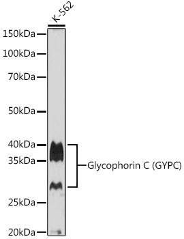
Western blot analysis of lysates from K-562 cells, using Glycophorin C (GYPC) Rabbit mAb (CAB11472) at 1:1000 dilution. Secondary antibody: HRP-conjugated Goat anti-Rabbit IgG (H+L) (CABS014) at 1:10000 dilution. Lysates/proteins: 25μg per lane. Blocking buffer: 3% nonfat dry milk in TBST. Detection: ECL Basic Kit (AbGn00020). Exposure time: 90s.
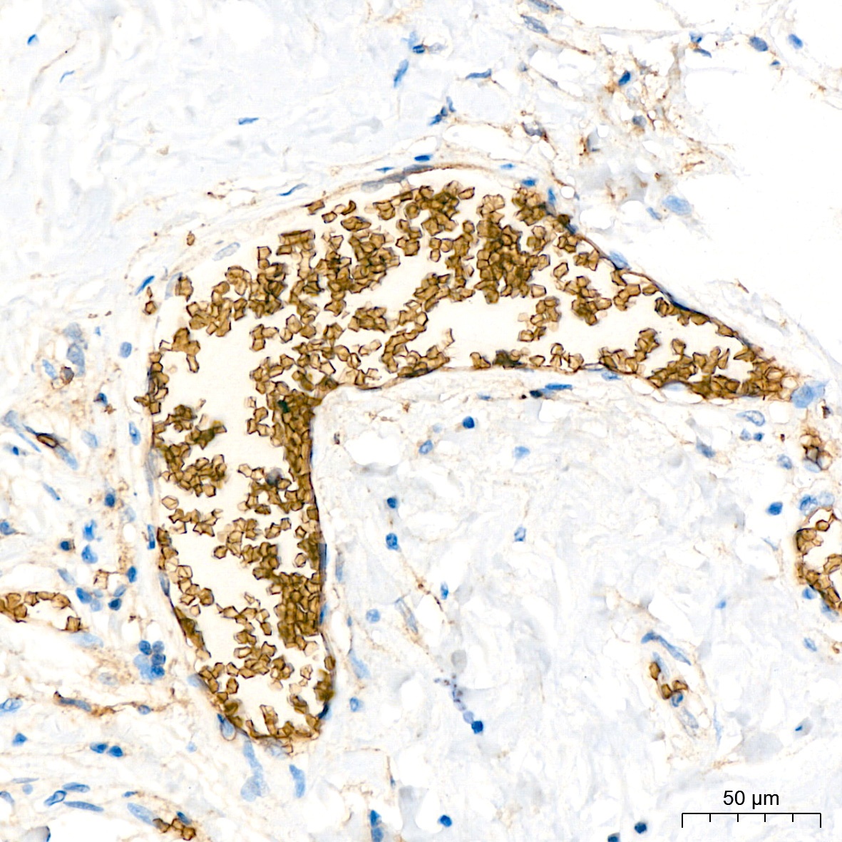
Immunohistochemistry analysis of paraffin-embedded Human breast tissue using Glycophorin C (GYPC) Rabbit mAb (CAB11472) at a dilution of 1:200 (40x lens). High pressure antigen retrieval performed with 0.01M Citrate buffer (pH 6.0) prior to IHC staining.
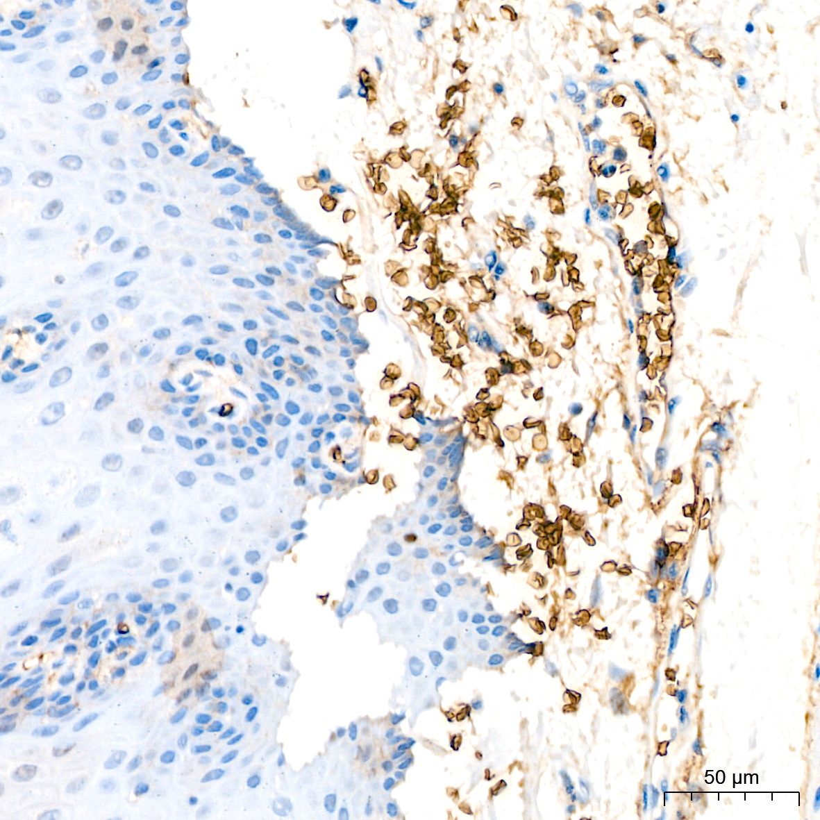
Immunohistochemistry analysis of paraffin-embedded Human esophagus tissue using Glycophorin C (GYPC) Rabbit mAb (CAB11472) at a dilution of 1:200 (40x lens). High pressure antigen retrieval performed with 0.01M Citrate buffer (pH 6.0) prior to IHC staining.
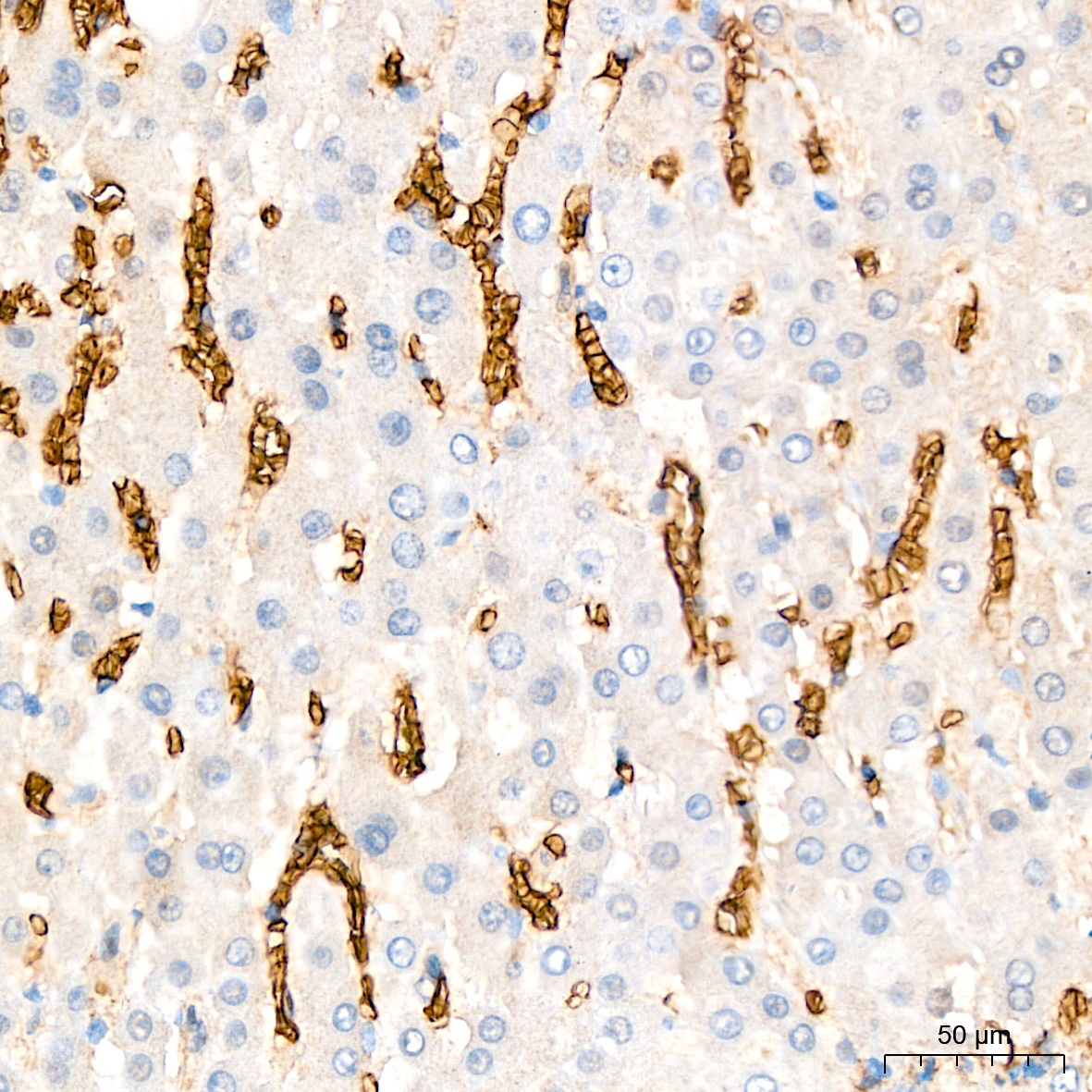
Immunohistochemistry analysis of paraffin-embedded Human liver tissue using Glycophorin C (GYPC) Rabbit mAb (CAB11472) at a dilution of 1:200 (40x lens). High pressure antigen retrieval performed with 0.01M Citrate buffer (pH 6.0) prior to IHC staining.