The HDAC3 Monoclonal Antibody (CAB19537) is a high-quality antibody developed for reliable detection and analysis of target proteins. This antibody, sourced from rabbits, is highly specific to HDAC3 and has been validated for use in a variety of applications, including Western blotting and immunofluorescence.HDAC3 plays a key role in diverse biological processes, such as cell proliferation, differentiation, and apoptosis, making it a target of interest in cancer research and developmental biology.
This antibody is validated for use in WB, IP, ELISA applications and has demonstrated reactivity against Human, Mouse, Rat samples.
Product Name:
HDAC3 Monoclonal Antibody
SKU:
CAB19537
Size:
20μL, 100μL
Reactivity:
Human, Mouse, Rat
Clone Number:
ARC0016
Conjugate:
Unconjugated
Immunogen:
Synthetic peptide. This information is considered to be commercially sensitive.
0.5μg-4μg antibody for 200μg-400μg extracts of whole cells
ELISA
Recommended starting concentration is 1 μg/mL. Please optimize the concentration based on your specific assay requirements.
Synonyms:
HD3, RPD3, KDAC3, RPD3-2, HDAC3
Positive Sample:
PC-3, HeLa, Mouse brain, NIH/3T3, Rat testis, 293T
Cellular Localization:
Cytoplasm, Nucleus, Cytosol.
Calculated MW:
49kDa
Observed MW:
49kDa
Histones play a critical role in transcriptional regulation, cell cycle progression, and developmental events. Histone acetylation/deacetylation alters chromosome structure and affects transcription factor access to DNA. The protein encoded by this gene belongs to the histone deacetylase/acuc/apha family. It has histone deacetylase activity and represses transcription when tethered to a promoter. It may participate in the regulation of transcription through its binding with the zinc-finger transcription factor YY1. This protein can also down-regulate p53 function and thus modulate cell growth and apoptosis. This gene is regarded as a potential tumor suppressor gene.
Purification Method
Affinity purification
Gene ID
8841
RRID
AB_2862654
Buffer Information
Store at -20℃. Avoid freeze / thaw cycles. Buffer: PBS containing 50% glycerol and 0.05% BSA, preserved with proclin300 or sodium azide, pH 7.3.
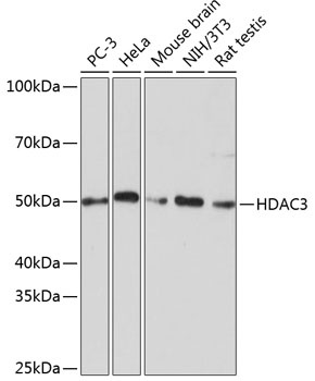
Western blot analysis of various lysates using [KD Validated] HDAC3 Rabbit mAb (CAB19537) at 1:1000 dilution incubated overnight at 4℃. Secondary antibody: HRP-conjugated Goat anti-Rabbit IgG (H+L) (CABS014) at 1:10000 dilution. Lysates/proteins: 25μg per lane. Blocking buffer: 3% nonfat dry milk in TBST. Detection: ECL Basic Kit (AbGn00020). Exposure time: 1min.
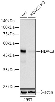
Western blot analysis of lysates from wild type (WT) and HDAC3 knockdown (KD) 293T cells using [KD Validated] HDAC3 Rabbit mAb (CAB19537) at 1:1000 dilution incubated overnight at 4℃. Secondary antibody: HRP-conjugated Goat anti-Rabbit IgG (H+L) (CABS014) at 1:10000 dilution. Lysates/proteins: 25 μg per lane. Blocking buffer: 3% nonfat dry milk in TBST. Detection: ECL Basic Kit (AbGn00020). Exposure time: 30s.


![Anti-HDAC3 [R03-4J9] Monoclonal Antibody (AGMB02156)](https://cdn11.bigcommerce.com/s-h68l9z2lnx/images/stencil/590x590/products/273445/678345/anti-hdac3-r03-4j9-monoclonal-antibody-agmb02156__76764.1773035436.jpg?c=2 "Anti-HDAC3 [R03-4J9] Monoclonal Antibody (AGMB02156)")
![Anti-HDAC3 [R07-8E9] Monoclonal Antibody (AGMB02801)](https://cdn11.bigcommerce.com/s-h68l9z2lnx/images/stencil/590x590/products/274090/678795/anti-hdac3-r07-8e9-monoclonal-antibody-agmb02801__74889.1773036871.jpg?c=2 "Anti-HDAC3 [R07-8E9] Monoclonal Antibody (AGMB02801)")

![Anti-HDAC3 (3G3) [3G3-H6-H10] Monoclonal Antibody (AGMB04528)](https://cdn11.bigcommerce.com/s-h68l9z2lnx/images/stencil/590x590/products/275816/676540/anti-hdac3-3g3-3g3-h6-h10-monoclonal-antibody-agmb04528__36996.1773029778.jpg?c=2 "Anti-HDAC3 (3G3) [3G3-H6-H10] Monoclonal Antibody (AGMB04528)")