The IMPDH2 Monoclonal Antibody (CAB9208) is a high-quality antibody developed for reliable detection and analysis of target proteins. This antibody, produced in rabbits, has been validated for use in Western blotting and immunohistochemistry applications with human samples.IMPDH2 plays a crucial role in cell proliferation and is upregulated in various types of cancer, making it a promising therapeutic target. This monoclonal antibody binds specifically to IMPDH2, allowing for precise detection and analysis in different cell types, making it an essential tool for studies in cancer biology and drug development.
This antibody is validated for use in WB, IF/ICC, ELISA applications and has demonstrated reactivity against Human, Mouse, Rat samples.
Product Name:
IMPDH2 Monoclonal Antibody
SKU:
CAB9208
Size:
20μL, 100μL
Reactivity:
Human, Mouse, Rat
Clone Number:
ARC1479
Conjugate:
Unconjugated
Immunogen:
Recombinant protein (or fragment).This information is considered to be commercially sensitive.
Recommended starting concentration is 1 μg/mL. Please optimize the concentration based on your specific assay requirements.
Synonyms:
IMPD2, IMPDH-II, H2
Positive Sample:
HeLa, K-562, NIH/3T3
Cellular Localization:
Cytoplasm, Nucleus.
Calculated MW:
56kDa
Observed MW:
56kDa
This gene encodes the rate-limiting enzyme in the de novo guanine nucleotide biosynthesis. It is thus involved in maintaining cellular guanine deoxy- and ribonucleotide pools needed for DNA and RNA synthesis. The encoded protein catalyzes the NAD-dependent oxidation of inosine-5'-monophosphate into xanthine-5'-monophosphate, which is then converted into guanosine-5'-monophosphate. This gene is up-regulated in some neoplasms, suggesting it may play a role in malignant transformation.
Purification Method
Affinity purification
Gene ID
3615
RRID
AB_2863688
Buffer Information
Store at -20℃. Avoid freeze / thaw cycles. Buffer: PBS containing 50% glycerol and 0.05% BSA, preserved with proclin300 or sodium azide, pH 7.3.
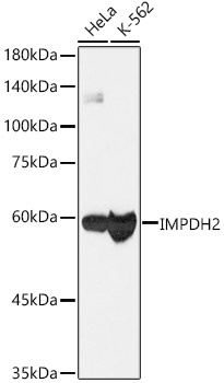
Western blot analysis of various lysates, using [KO Validated] IMPDH2 Rabbit mAb (CAB9208) at 1:1000 dilution. Secondary antibody: HRP-conjugated Goat anti-Rabbit IgG (H+L) (CABS014) at 1:10000 dilution. Lysates/proteins: 25μg per lane. Blocking buffer: 3% nonfat dry milk in TBST. Detection: ECL Basic Kit (AbGn00020). Exposure time: 3s.
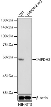
Western blot analysis of lysates from wild type(WT) and IMPDH2 knockout (KO) NIH/3T3(KO) cells, using [KO Validated] IMPDH2 Rabbit mAb (CAB9208) at 1:1000 dilution. Secondary antibody: HRP-conjugated Goat anti-Rabbit IgG (H+L) (CABS014) at 1:10000 dilution. Lysates/proteins: 25μg per lane. Blocking buffer: 3% nonfat dry milk in TBST. Detection: ECL Basic Kit (AbGn00020). Exposure time: 3s.
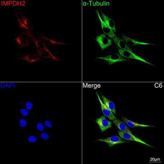
Confocal imaging of C6 cells using [KO Validated] IMPDH2 Rabbit mAb (CAB9208, dilution 1:200) followed by a further incubation with Cy3 Goat Anti-Rabbit IgG (H+L) (CABS007, dilution 1:500) (Red). The cells were counterstained with α-Tubulin Mouse mAb (AC012, dilution 1:400) followed by incubation with ABflo® 488-conjugated Goat Anti-Mouse IgG (H+L) Ab (CABS076, dilution 1:500) (Green). DAPI was used for nuclear staining (Blue). Objective: 100x.
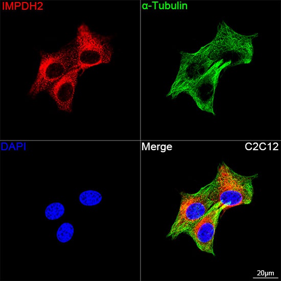
Confocal imaging of C2C12 cells using [KO Validated] IMPDH2 Rabbit mAb (CAB9208, dilution 1:200) followed by a further incubation with Cy3 Goat Anti-Rabbit IgG (H+L) (CABS007, dilution 1:500) (Red). The cells were counterstained with α-Tubulin Mouse mAb (AC012, dilution 1:400) followed by incubation with ABflo® 488-conjugated Goat Anti-Mouse IgG (H+L) Ab (CABS076, dilution 1:500) (Green). DAPI was used for nuclear staining (Blue). Objective: 100x.


![Anti-IMPDH2 [R06-1B8] Monoclonal Antibody (AGMB02504)](https://cdn11.bigcommerce.com/s-h68l9z2lnx/images/stencil/590x590/products/273793/678243/anti-impdh2-r06-1b8-monoclonal-antibody-agmb02504__26443.1773035175.jpg?c=2 "Anti-IMPDH2 [R06-1B8] Monoclonal Antibody (AGMB02504)")


![Anti-IMPDH2 [R07-1V9] Monoclonal Antibody (AGMB03097)](https://cdn11.bigcommerce.com/s-h68l9z2lnx/images/stencil/590x590/products/274386/677920/anti-impdh2-r07-1v9-monoclonal-antibody-agmb03097__26342.1773034110.jpg?c=2 "Anti-IMPDH2 [R07-1V9] Monoclonal Antibody (AGMB03097)")

