The [KO Validated] Cyclin D1 Monoclonal Antibody (CAB19038) is a high-quality antibody developed for reliable detection and analysis of target proteins. The protein encoded by this gene belongs to the highly conserved cyclin family, whose members are characterized by a dramatic periodicity in protein abundance throughout the cell cycle. Cyclins function as regulators of CDK kinases. Different cyclins exhibit distinct expression and degradation patterns which contribute to the temporal coordination of each mitotic event. This cyclin forms a complex with and functions as a regulatory subunit of CDK4 or CDK6, whose activity is required for cell cycle G1/S transition. This protein has been shown to interact with tumor suppressor protein Rb and the expression of this gene is regulated positively by Rb. Mutations, amplification and overexpression of this gene, which alters cell cycle progression, are observed frequently in a variety of human cancers. RRID AB_2862530 Gene ID 595 Swiss Prot Synonym BCL1; PRAD1; U21B31; D11S287E; D1
This antibody is validated for use in WB, IF/ICC, ELISA applications and has demonstrated reactivity against Human, Mouse, Rat samples.
Product Name:
[KO Validated] Cyclin D1 Monoclonal Antibody
SKU:
CAB19038
Size:
100μL, 20μL
Reactivity:
Human, Mouse, Rat
Clone Number:
ARC0300
Conjugate:
Unconjugated
Immunogen:
Synthetic peptide. This information is considered to be commercially sensitive.
Tested Applications:
WBIF/ICCELISA
Recommended Dilution:
WB
1:1000 - 1:5000
IF
/
ICC
1:100 - 1:800
ELISA
Recommended starting concentration is 1 μg/mL. Please optimize the concentration based on your specific assay requirements.
Synonyms:
BCL1, PRAD1, U21B31, D11S287E, D1
Positive Sample:
Hep G2, MCF7, HeLa, Mouse lung, C6
Cellular Localization:
Nucleus.
Calculated MW:
34kDa
Observed MW:
34kDa/36kDa
The protein encoded by this gene belongs to the highly conserved cyclin family, whose members are characterized by a dramatic periodicity in protein abundance throughout the cell cycle. Cyclins function as regulators of CDK kinases. Different cyclins exhibit distinct expression and degradation patterns which contribute to the temporal coordination of each mitotic event. This cyclin forms a complex with and functions as a regulatory subunit of CDK4 or CDK6, whose activity is required for cell cycle G1/S transition. This protein has been shown to interact with tumor suppressor protein Rb and the expression of this gene is regulated positively by Rb. Mutations, amplification and overexpression of this gene, which alters cell cycle progression, are observed frequently in a variety of human cancers. RRID AB_2862530 Gene ID 595 Swiss Prot Synonym BCL1; PRAD1; U21B31; D11S287E; D1
Purification Method:
Affinity purification
Gene ID:
595
RRID:
AB_2862530
Buffer Information:
Store at -20℃. Avoid freeze / thaw cycles. Buffer: PBS containing 50% glycerol and 0.05% BSA, preserved with proclin300 or sodium azide, pH 7.3.
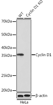
Western blot analysis of lysates from wild type (WT) and Cyclin D1 knockout (KO) HeLa cells, using [KO Validated] Cyclin D1 Rabbit mAb (CAB19038) at 1:1000 dilution. Secondary antibody: HRP-conjugated Goat anti-Rabbit IgG (H+L) (AS014) at 1:10000 dilution. Lysates/proteins: 25μg per lane. Blocking buffer: 3% nonfat dry milk in TBST. Detection: ECL Basic Kit (AbGn00020). Exposure time: 1min.
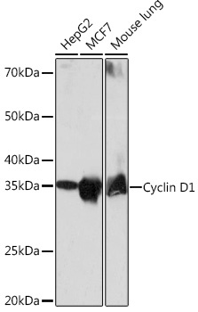
Western blot analysis of various lysates using [KO Validated] Cyclin D1 Rabbit mAb (CAB19038) at 1:1000 dilution. Secondary antibody: HRP-conjugated Goat anti-Rabbit IgG (H+L) (AS014) at 1:10000 dilution. Lysates/proteins: 25μg per lane. Blocking buffer: 3% nonfat dry milk in TBST. Detection: ECL Basic Kit (AbGn00020). Exposure time: 1min.
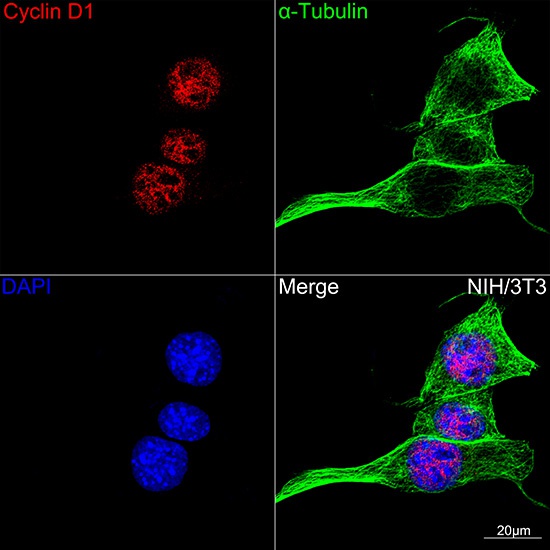
Confocal imaging of NIH/3T3 cells using [KO Validated] Cyclin D1 Rabbit mAb (CAB19038,dilution 1:100)(Red). The cells were counterstained with α-Tubulin Mouse mAb (AC012,dilution 1:400) (Green). DAPI was used for nuclear staining (blue). Objective: 100x.
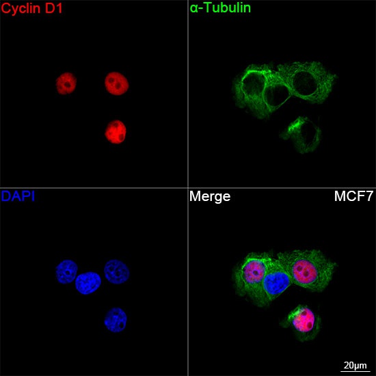
Confocal imaging of MCF7 cells using [KO Validated] Cyclin D1 Rabbit mAb (CAB19038, dilution 1:200) followed by a further incubation with Cy3 Goat Anti-Rabbit IgG (H+L) (AS007, dilution 1:500) (Red). The cells were counterstained with α-Tubulin Mouse mAb (AC012, dilution 1:400) followed by incubation with ABflo® 488-conjugated Goat Anti-Mouse IgG (H+L) Ab (AS076, dilution 1:500) (Green). DAPI was used for nuclear staining (Blue). Objective: 100x.