The [KO Validated] HK1 Monoclonal Antibody (CAB0533) is a high-quality antibody developed for reliable detection and analysis of target proteins. This antibody, produced using rabbits, has been validated for use in various applications, including Western blot and immunohistochemistry. Its high specificity and sensitivity make it ideal for detecting and analyzing HK1 protein levels in biological samples.HK1 is a key enzyme in the glycolytic pathway and is overexpressed in many cancer types, contributing to the high energy demands of rapidly proliferating tumor cells.
This antibody is validated for use in WB, IHC-P, IF/ICC, IP, ELISA, IF-P applications and has demonstrated reactivity against Human, Mouse, Rat samples.
Product Name:
[KO Validated] HK1 Monoclonal Antibody
SKU:
CAB0533
Size:
20μL, 100μL
Reactivity:
Human, Mouse, Rat
Clone Number:
ARC0256
Conjugate:
Unconjugated
Immunogen:
Synthetic peptide. This information is considered to be commercially sensitive.
293T, MCF7, Mouse testis, Mouse brain, Rat testis, Rat brain
Cellular Localization:
Mitochondrion Outer Membrane.
Calculated MW:
102kDa
Observed MW:
120kDa
Hexokinases phosphorylate glucose to produce glucose-6-phosphate, the first step in most glucose metabolism pathways. This gene encodes a ubiquitous form of hexokinase which localizes to the outer membrane of mitochondria. Mutations in this gene have been associated with hemolytic anemia due to hexokinase deficiency. Alternative splicing of this gene results in several transcript variants which encode different isoforms, some of which are tissue-specific.
Purification Method
Affinity purification
Gene ID
3098
RRID
AB_2861463
Buffer Information
Store at -20℃. Avoid freeze / thaw cycles. Buffer: PBS containing 50% glycerol and 0.05% BSA, preserved with proclin300 or sodium azide, pH 7.3.
Western blot analysis of lysates from wild type (WT) and HK1 knockout (KO) 293T cells, using [KO Validated] HK1 Rabbit mAb (CAB0533) at 1:1000 dilution. Secondary antibody: HRP-conjugated Goat anti-Rabbit IgG (H+L) (CABS014) at 1:10000 dilution. Lysates/proteins: 25μg per lane. Blocking buffer: 3% nonfat dry milk in TBST. Detection: ECL Basic Kit (AbGn00020). Exposure time: 60s.
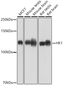
Western blot analysis of various lysates using [KO Validated] HK1 Rabbit mAb (CAB0533) at 1:1000 dilution. Secondary antibody: HRP-conjugated Goat anti-Rabbit IgG (H+L) (CABS014) at 1:10000 dilution. Lysates/proteins: 25μg per lane. Blocking buffer: 3% nonfat dry milk in TBST. Detection: ECL Basic Kit (AbGn00020). Exposure time: 60s.
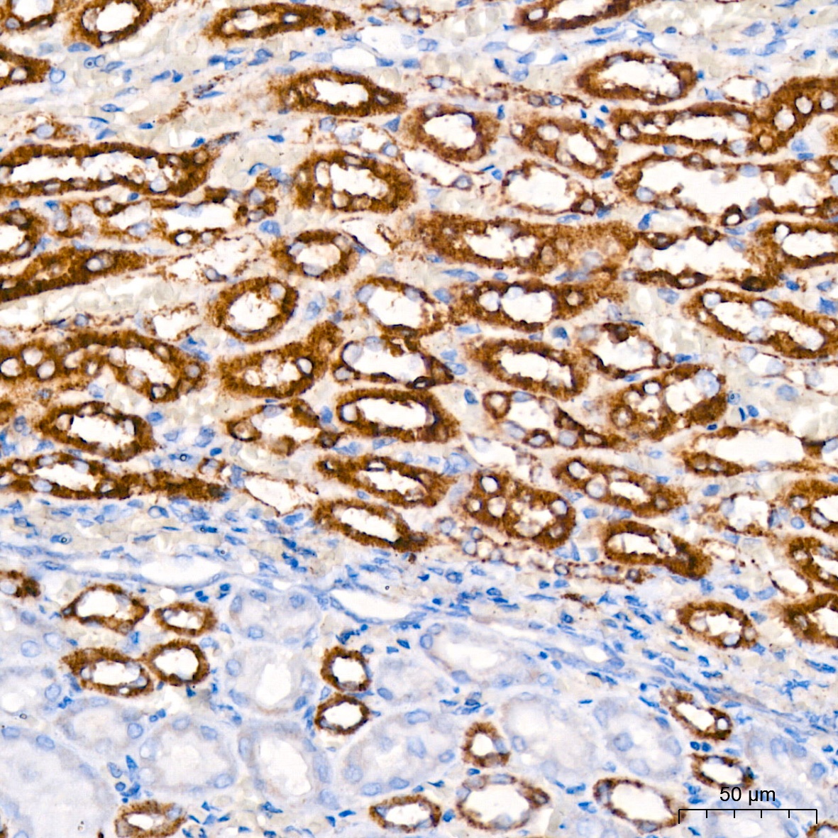
Immunohistochemistry analysis of paraffin-embedded Rat kidney tissue using [KO Validated] HK1 Rabbit mAb (CAB0533) at dilution of 1:200 (40x lens). High pressure antigen retrieval performed with 0.01M Citrate buffer (pH 6.0) prior to IHC staining.
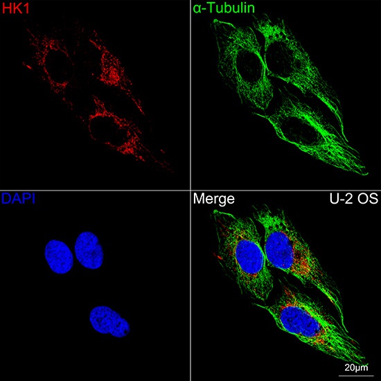
Confocal imaging of U-2 OS cells using [KO Validated] HK1 Rabbit mAb (CAB0533, dilution 1:200) followed by a further incubation with Cy3 Goat Anti-Rabbit IgG (H+L) (CABS007, dilution 1:500) (Red). The cells were counterstained with α-Tubulin Mouse mAb (AC012, dilution 1:400) followed by incubation with ABflo® 488-conjugated Goat Anti-Mouse IgG (H+L) Ab (CABS076, dilution 1:500) (Green). DAPI was used for nuclear staining (Blue). Objective: 100x.
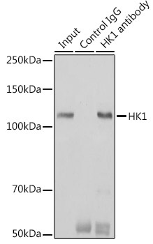
Immunoprecipitation analysis of 600 μg extracts of Mouse brain cells using 3 μg [KO Validated] HK1 Rabbit mAb (CAB0533). Western blot was performed from the immunoprecipitate using HK1 (CAB0533) at a dilution of 1:1000.