The [KO Validated] SQSTM1/p62 Monoclonal Antibody (CAB19700) is a high-quality antibody developed for reliable detection and analysis of target proteins. This antibody, produced in rabbits, exhibits high specificity and sensitivity towards SQSTM1/P62 in human samples, making it ideal for Western blot applications.SQSTM1/P62 is implicated in the maintenance of cellular homeostasis and plays a crucial role in the clearance of protein aggregates and damaged organelles through autophagy. Dysregulation of SQSTM1/P62 has been linked to various diseases including neurodegenerative disorders, cancer, and metabolic diseases, making it a promising target for therapeutic interventions.
This antibody is validated for use in WB, IHC-P, IF/ICC, IP, ELISA applications and has demonstrated reactivity against Human, Mouse, Rat samples.
Product Name:
[KO Validated] SQSTM1/p62 Monoclonal Antibody
SKU:
CAB19700
Size:
20μL, 100μL
Reactivity:
Human, Mouse, Rat
Clone Number:
ARC0180
Conjugate:
Unconjugated
Immunogen:
Synthetic peptide. This information is considered to be commercially sensitive.
This gene encodes a multifunctional protein that binds ubiquitin and regulates activation of the nuclear factor kappa-B (NF-kB) signaling pathway. The protein functions as a scaffolding/adaptor protein in concert with TNF receptor-associated factor 6 to mediate activation of NF-kB in response to upstream signals. Alternatively spliced transcript variants encoding either the same or different isoforms have been identified for this gene. Mutations in this gene result in sporadic and familial Paget disease of bone.
Purification Method
Affinity purification
Gene ID
8878
RRID
AB_2862742
Buffer Information
Store at -20℃. Avoid freeze / thaw cycles. Buffer: PBS containing 50% glycerol and 0.05% BSA, preserved with proclin300 or sodium azide, pH 7.3.
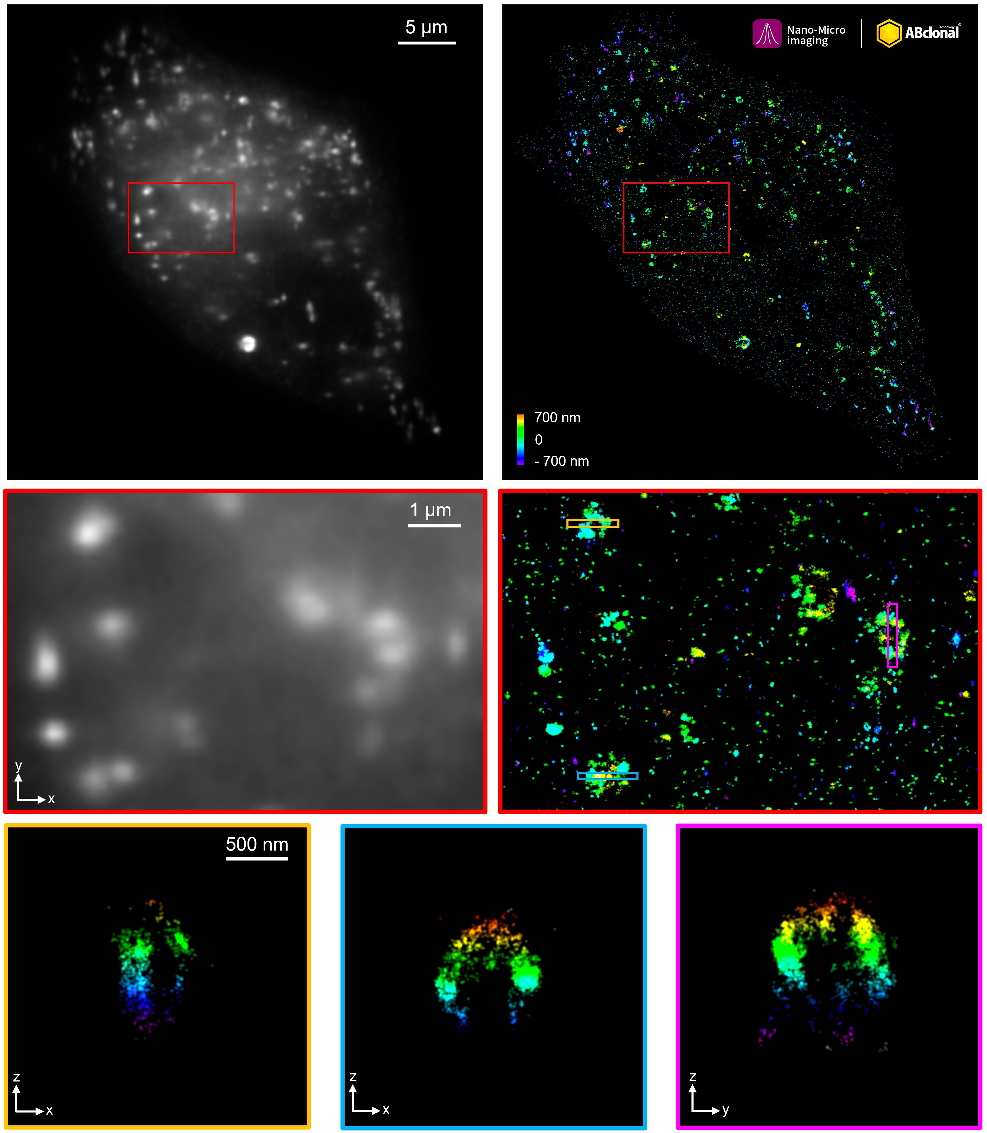
The STORM super-resolution (SR) imaging of HeLa cells using SQSTM1/p62 Rabbit mAb (CAB19700, ABclonal) at dilution of 1:200 with 3% paraformaldehyde (PFA) +0.1% glutaraldehyde (GA) fixation. The immunostaining was performed by Full Automatic Immunofluorescence Workflow System (Workflow Ultra300, Nano-Micro imaging, China). Image was performed with Single-Molecule Localization Super-Resolution Microscopy (STORM Ultra300, Nano-Micro imaging, China). We acknowledge Nano-Micro imaging Biotechnology Co., Ltd. in SR image processing and kindly providing this image.
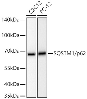
Western blot analysis of various lysates using SQSTM1/p62 Rabbit mAb (CAB19700) at 1:20000 dilution incubated overnight at 4℃. Secondary antibody: HRP-conjugated Goat anti-Rabbit IgG (H+L) (CABS014) at 1:10000 dilution. Lysates/proteins: 25 μg per lane. Blocking buffer: 3% nonfat dry milk in TBST. Detection: ECL Basic Kit (AbGn00020). Exposure time: 10s.
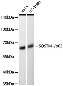
Western blot analysis of various lysates using SQSTM1/p62 Rabbit mAb (CAB19700) at 1:20000 dilution incubated overnight at 4℃. Secondary antibody: HRP-conjugated Goat anti-Rabbit IgG (H+L) (CABS014) at 1:10000 dilution. Lysates/proteins: 25 μg per lane. Blocking buffer: 3% nonfat dry milk in TBST. Detection: ECL Basic Kit (AbGn00020). Exposure time: 30s.
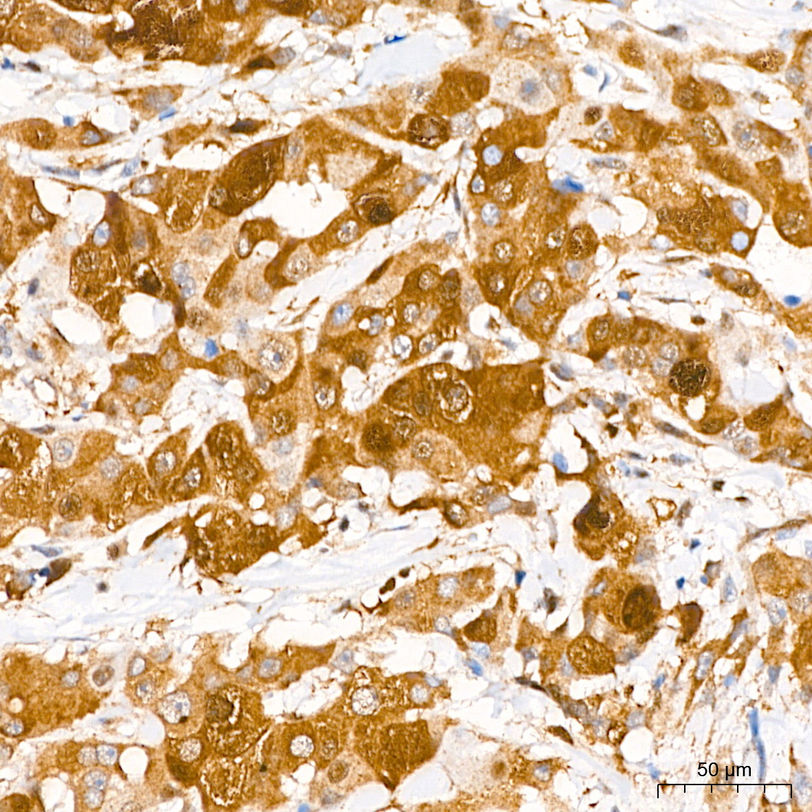
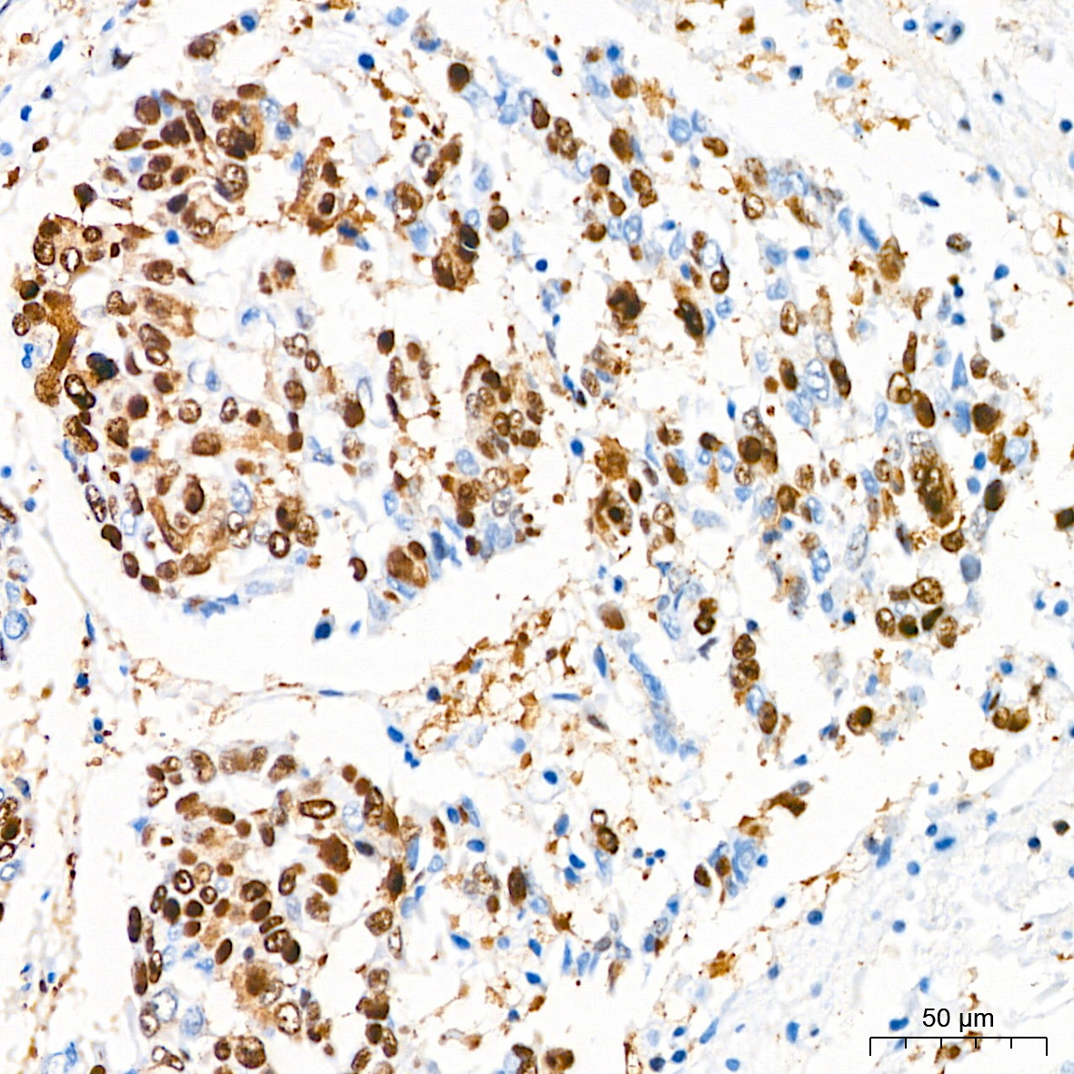
Immunohistochemistry analysis of paraffin-embedded Human breast cancer tissue using SQSTM1/p62 Rabbit mAb (CAB19700) at a dilution of 1:500 (40x lens). High pressure antigen retrieval performed with 0.01M Citrate Buffer(pH 6.0) prior to IHC staining.
Immunohistochemistry analysis of paraffin-embedded Human colon tissue using SQSTM1/p62 Rabbit mAb (CAB19700) at a dilution of 1:500 (40x lens). High pressure antigen retrieval performed with 0.01M Citrate Buffer(pH 6.0) prior to IHC staining.
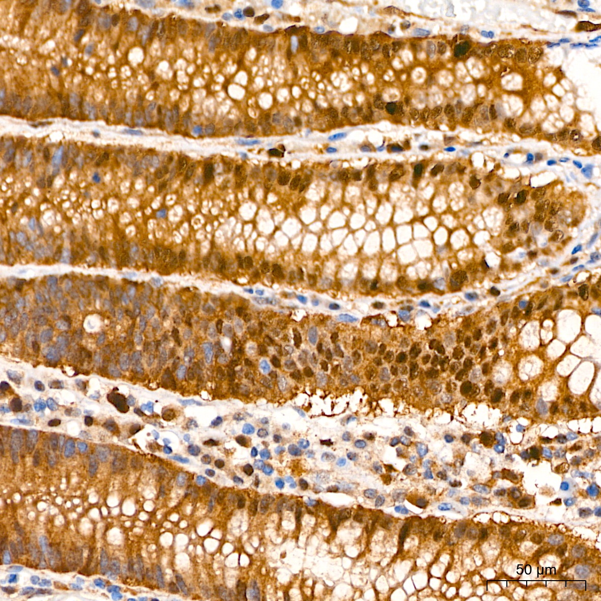
Immunohistochemistry analysis of paraffin-embedded Human esophagus tissue using SQSTM1/p62 Rabbit mAb (CAB19700) at a dilution of 1:500 (40x lens). High pressure antigen retrieval performed with 0.01M Citrate Buffer(pH 6.0) prior to IHC staining.
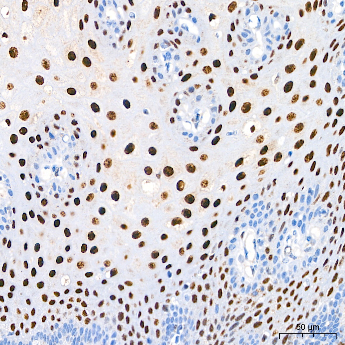
Immunohistochemistry analysis of paraffin-embedded Human lung squamous carcinoma tissue using SQSTM1/p62 Rabbit mAb (CAB19700) at a dilution of 1:500 (40x lens). High pressure antigen retrieval performed with 0.01M Citrate Buffer(pH 6.0) prior to IHC staining.
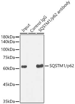
Immunoprecipitation analysis of 300 μg extracts from HeLa cells using 3 μg SQSTM1/p62 Rabbit mAb (CAB19700). Western blot was performed from the immunoprecipitate using SQSTM1/p62 Rabbit mAb (CAB19700) at a dilition of 1:1000.