The LRAT Antibody (CAB13256) is a high-quality antibody developed for reliable detection and analysis of target proteins. The antibody, generated in rabbits, exhibits high reactivity with human samples and is validated for use in Western blot applications. By binding specifically to the LRAT protein, this antibody enables precise detection and analysis in a variety of cell types, making it an essential component for investigations in the fields of biochemistry and nutrition.LRAT plays a crucial role in the conversion of retinol (vitamin A) into retinyl esters, which are necessary for normal vision and overall health.
This antibody is validated for use in WB, IF/ICC, ELISA applications and has demonstrated reactivity against Human, Mouse, Rat samples.
Product Name:
LRAT Antibody
SKU:
CAB13256
Size:
20μL, 100μL
Reactivity:
Human, Mouse, Rat
Conjugate:
Unconjugated
Immunogen:
Recombinant protein (or fragment).This information is considered to be commercially sensitive.
The protein encoded by this gene localizes to the endoplasmic reticulum, where it catalyzes the esterification of all-trans-retinol into all-trans-retinyl ester. This reaction is an important step in vitamin A metabolism in the visual system. Mutations in this gene have been associated with early-onset severe retinal dystrophy and Leber congenital amaurosis 14. Alternative splicing results in multiple transcript variants.
Purification Method
Affinity purification
Gene ID
9227
RRID
AB_2760109
Buffer Information
Store at -20℃. Avoid freeze / thaw cycles. Buffer: PBS containing 50% glycerol, preserved with proclin300 or sodium azide, pH 7.3.
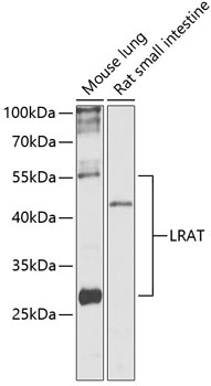
Western blot analysis of various lysates using LRAT Rabbit pAb (CAB13256) at 1:1000 dilution. Secondary antibody: HRP-conjugated Goat anti-Rabbit IgG (H+L) (CABS014) at 1:10000 dilution. Lysates/proteins: 25μg per lane. Blocking buffer: 3% nonfat dry milk in TBST. Detection: ECL Basic Kit (AbGn00020). Exposure time: 30s.
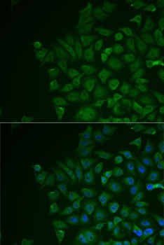
Immunofluorescence analysis of MCF7 cells using LRAT Rabbit pAb (CAB13256). Secondary antibody: Cy3-conjugated Goat anti-Rabbit IgG (H+L) (CABS007) at 1:500 dilution. Blue: DAPI for nuclear staining.