The MELK Antibody (CAB10794) is a high-quality antibody developed for reliable detection and analysis of target proteins. This antibody, developed in rabbits, exhibits high reactivity with human samples and has been validated for use in Western blot applications. By binding to the MELK protein, this antibody enables precise detection and analysis in various cell types, making it an essential resource for studies in cancer biology and therapeutic development.
This antibody is validated for use in WB, ELISA applications and has demonstrated reactivity against Human, Mouse, Rat samples.
Product Name:
MELK Antibody
SKU:
CAB10794
Size:
20μL, 100μL
Reactivity:
Human, Mouse, Rat
Conjugate:
Unconjugated
Immunogen:
Recombinant protein (or fragment).This information is considered to be commercially sensitive.
Recommended starting concentration is 1 μg/mL. Please optimize the concentration based on your specific assay requirements.
Synonyms:
HPK38, MELK
Positive Sample:
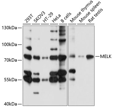
293T, SKOV3, HT-29, HeLa, B cells, Mouse thymus, Mouse spleen, Rat testis
Cellular Localization:
Cell Membrane, Peripheral Membrane Protein.
Calculated MW:
75kDa
Observed MW:
75kDa
Enables calcium ion binding activity; non-membrane spanning protein tyrosine kinase activity; and protein serine/threonine kinase activity. Involved in apoptotic process; cell population proliferation; and protein autophosphorylation. Located in cell cortex and plasma membrane.
Purification Method
Affinity purification
Gene ID
9833
RRID
AB_2758228
Buffer Information
Store at -20℃. Avoid freeze / thaw cycles. Buffer: PBS with 0.01% thimerosal,50% glycerol,pH7.3.
Western blot analysis of various lysates using MELK Rabbit pAb (CAB10794) at 1:1000 dilution. Secondary antibody: HRP-conjugated Goat anti-Rabbit IgG (H+L) (CABS014) at 1:10000 dilution. Lysates/proteins: 25μg per lane. Blocking buffer: 3% nonfat dry milk in TBST. Detection: ECL Basic Kit (AbGn00020). Exposure time: 5s.