The [KO Validated] PD-1/CD279 Polyclonal Antibody (CAB5584) is a high-quality antibody developed for reliable detection and analysis of target proteins. This cell surface receptor is a critical regulator of immune responses, particularly in the context of immune tolerance and the prevention of autoimmune diseases. Raised in rabbits, this antibody exhibits high specificity towards human samples and has been validated for Western blot applications. By binding to the PDCD1 protein, researchers can accurately detect and analyze its expression in various cell types, making it an invaluable asset for studies in immunology, oncology, and autoimmune disorders.
This antibody is validated for use in WB, ELISA applications and has demonstrated reactivity against Human, Mouse, Rat samples.
Product Name:
[KO Validated] PD-1/CD279 Polyclonal Antibody
SKU:
CAB5584
Size:
20μL, 100μL
Reactivity:
Human, Mouse, Rat
Conjugate:
Unconjugated
Immunogen:
Recombinant protein (or fragment).This information is considered to be commercially sensitive.
Recommended starting concentration is 1 μg/mL. Please optimize the concentration based on your specific assay requirements.
Synonyms:
PD1, PD-1, CD279, SLEB2, hPD-1, hPD-l, hSLE1, 79
Positive Sample:
293T, RAW264.7, Mouse spleen, Mouse thymus, Rat spleen, Rat thymus
Cellular Localization:
Membrane, Single-Pass Type I Membrane Protein.
Calculated MW:
32kDa
Observed MW:
35kDa
Programmed cell death protein 1 (PDCD1) is an immune-inhibitory receptor expressed in activated T cells; it is involved in the regulation of T-cell functions, including those of effector CD8+ T cells. In addition, this protein can also promote the differentiation of CD4+ T cells into T regulatory cells. PDCD1 is expressed in many types of tumors including melanomas, and has demonstrated to play a role in anti-tumor immunity. Moreover, this protein has been shown to be involved in safeguarding against autoimmunity, however, it can also contribute to the inhibition of effective anti-tumor and anti-microbial immunity.
Purification Method
Affinity purification
Gene ID
5133
RRID
AB_2766357
Buffer Information
Store at -20℃. Avoid freeze / thaw cycles. Buffer: PBS containing 50% glycerol, preserved with proclin300 or sodium azide, pH 7.3.
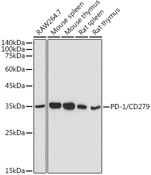
Western blot analysis of various lysates using [KO Validated] PD-1/CD279 Rabbit pAb (CAB5584) at 1:500 dilution. Secondary antibody: HRP-conjugated Goat anti-Rabbit IgG (H+L) (CABS014) at 1:10000 dilution. Lysates/proteins: 25μg per lane. Blocking buffer: 3% nonfat dry milk in TBST. Detection: ECL Basic Kit (AbGn00020). Exposure time: 90s.
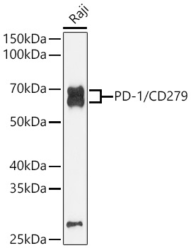
Western blot analysis of lysates from Raji cells using PD-1/CD279 Rabbit pAb (CAB5584) at 1:1000 dilution. Secondary antibody: HRP-conjugated Goat anti-Rabbit IgG (H+L) (CABS014) at 1:10000 dilution. Lysates/proteins: 25 μg per lane. Blocking buffer: 3% nonfat dry milk in TBST. Detection: ECL Enhanced Kit (AbGn00021). Exposure time: 180s.