The PDK4 Antibody (CAB13337) is a high-quality antibody developed for reliable detection and analysis of target proteins. This antibody, produced in rabbits, exhibits high specificity and sensitivity towards human samples, making it ideal for use in Western blot applications.PDK4 is known to play a crucial role in the regulation of glucose and fatty acid metabolism, making it a potential target for research in metabolic disorders such as diabetes and obesity. By targeting the PDK4 protein, researchers can gain insights into the mechanisms underlying metabolic dysfunction and identify potential therapeutic strategies for these conditions.
This antibody is validated for use in WB, IHC-P, IF/ICC, ELISA applications and has demonstrated reactivity against Human, Mouse, Rat samples.
Product Name:
PDK4 Antibody
SKU:
CAB13337
Size:
20μL, 100μL
Reactivity:
Human, Mouse, Rat
Conjugate:
Unconjugated
Immunogen:
Synthetic peptide. This information is considered to be commercially sensitive.
Recommended starting concentration is 1 μg/mL. Please optimize the concentration based on your specific assay requirements.
Synonyms:
PDK4
Positive Sample:
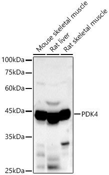
Mouse skeletal muscle, Rat liver, Rat skeletal muscle
Cellular Localization:
Mitochondrion Matrix.
Calculated MW:
46kDa
Observed MW:
46kDa
This gene is a member of the PDK/BCKDK protein kinase family and encodes a mitochondrial protein with a histidine kinase domain. This protein is located in the matrix of the mitrochondria and inhibits the pyruvate dehydrogenase complex by phosphorylating one of its subunits, thereby contributing to the regulation of glucose metabolism. Expression of this gene is regulated by glucocorticoids, retinoic acid and insulin.
Purification Method
Affinity purification
Gene ID
5166
RRID
AB_2760193
Buffer Information
Store at -20℃. Avoid freeze / thaw cycles. Buffer: PBS containing 50% glycerol, preserved with proclin300 or sodium azide, pH 7.3.
Western blot analysis of various lysates, using PDK4 Rabbit pAb (CAB13337) at 1:2000 dilution. Secondary antibody: HRP-conjugated Goat anti-Rabbit IgG (H+L) (CABS014) at 1:10000 dilution. Lysates/proteins: 25μg per lane. Blocking buffer: 3% nonfat dry milk in TBST. Detection: ECL Basic Kit (AbGn00020). Exposure time: 90s.
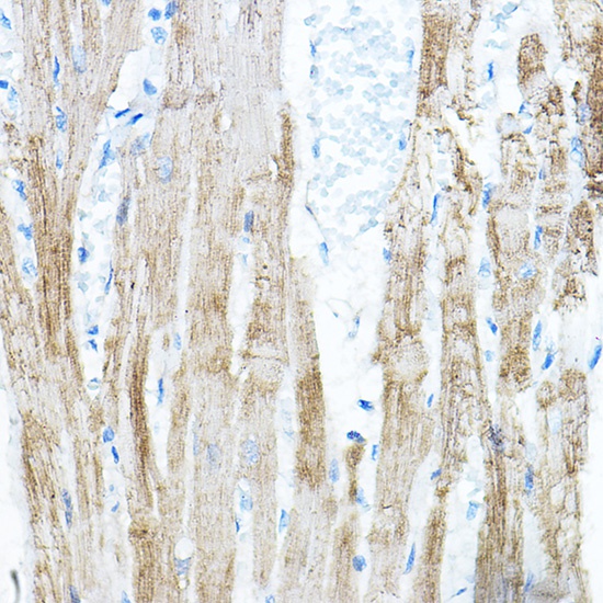
Immunohistochemistry analysis of paraffin-embedded Rat heart using PDK4 Rabbit pAb (CAB13337) at dilution of 1:100 (40x lens). High pressure antigen retrieval performed with 0.01M Citrate buffer (pH 6.0) prior to IHC staining.
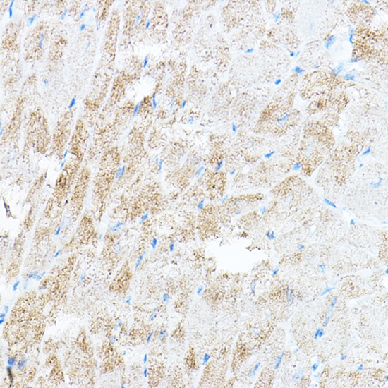
Immunohistochemistry analysis of paraffin-embedded Mouse heart using PDK4 Rabbit pAb (CAB13337) at dilution of 1:100 (40x lens). High pressure antigen retrieval performed with 0.01M Citrate buffer (pH 6.0) prior to IHC staining.
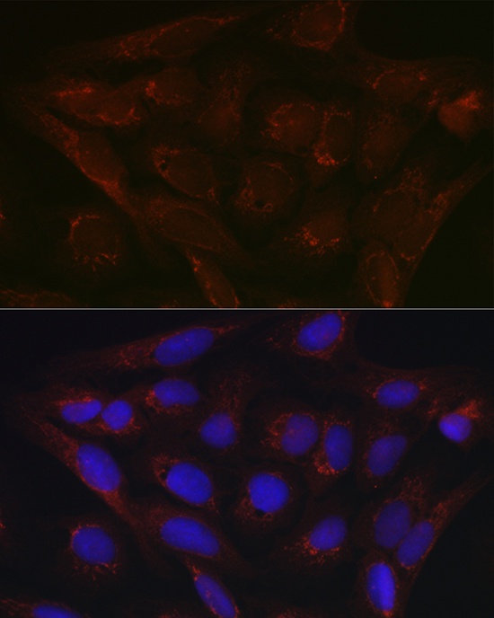
Immunofluorescence analysis of U2OS cells using PDK4 Rabbit pAb (CAB13337) at dilution of 1:100 (40x lens). Secondary antibody: Cy3-conjugated Goat anti-Rabbit IgG (H+L) (CABS007) at 1:500 dilution. Blue: DAPI for nuclear staining.