The Phospho-CDK2-Y15 Monoclonal Antibody (CABP1005) is a high-quality antibody developed for reliable detection and analysis of target proteins. This antibody specifically targets phosphorylated CDK2 at Tyrosine 15, a key regulatory site that controls the activity of this cyclin-dependent kinase. CDK2 is a crucial player in cell cycle progression, regulating entry into S phase and promoting cell division. Phosphorylation at Y15 inhibits CDK2 activity, preventing premature cell cycle progression and ensuring proper cell division. Dysregulation of CDK2 activity, often seen in cancer cells, can lead to uncontrolled cell proliferation and tumor formation.This antibody, generated in rabbits, is highly specific and sensitive, making it ideal for detecting changes in CDK2 phosphorylation in various cell types and experimental conditions.
This antibody is validated for use in WB, ELISA applications and has demonstrated reactivity against Human, Mouse samples.
Product Name:
Phospho-CDK2-Y15 Monoclonal Antibody
SKU:
CABP1005
Size:
20μL, 100μL
Reactivity:
Human, Mouse
Clone Number:
ARC1550
Conjugate:
Unconjugated
Immunogen:
Synthetic peptide. This information is considered to be commercially sensitive.
Sequence:
GTYG V
Tested Applications:
WBELISA
Recommended Dilution:
WB
1:1000 - 1:10000
ELISA
Recommended starting concentration is 1 μg/mL. Please optimize the concentration based on your specific assay requirements.
Synonyms:
CDKN2, p33(CDK2), Phospho-CDK2-Y15
Positive Sample:
NIH/3T3 treated with Hydroxyurea, NIH/3T3 treated with Nocodazole, HeLa treated with Hydroxyurea
This gene encodes a member of a family of serine/threonine protein kinases that participate in cell cycle regulation. The encoded protein is the catalytic subunit of the cyclin-dependent protein kinase complex, which regulates progression through the cell cycle. Activity of this protein is especially critical during the G1 to S phase transition. This protein associates with and regulated by other subunits of the complex including cyclin A or E, CDK inhibitor p21Cip1 (CDKN1A), and p27Kip1 (CDKN1B). Alternative splicing results in multiple transcript variants.
Purification Method
Affinity purification
Gene ID
1017
RRID
AB_2863897
Buffer Information
Store at -20℃. Avoid freeze / thaw cycles. Buffer: PBS containing 50% glycerol and 0.05% BSA, preserved with proclin300 or sodium azide, pH 7.3.
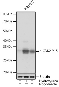
Western blot analysis of lysates from NIH/3T3 cells using Phospho-CDK2-Y15 Rabbit mAb (CABP1005) at 1:1000 dilution. NIH/3T3 cells were treated with Hydroxyurea (4 Mm) at 37℃ for 20 hours or treated with Nocodazole (100 ng/mL) at 37℃ for 24 hours. Secondary antibody: HRP-conjugated Goat anti-Rabbit IgG (H+L) (CABS014) at 1:10000 dilution. Lysates/proteins: 25μg per lane. Blocking buffer: 3% BSA. Detection: ECL Basic Kit (AbGn00020). Exposure time: 3 s.
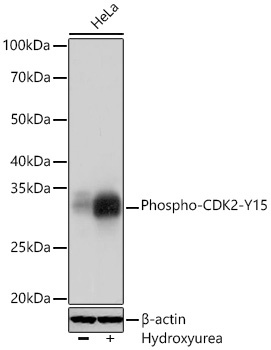
Western blot analysis of lysates from HeLa cells using Phospho-CDK2-Y15 Rabbit mAb (CABP1005) at 1:1000 dilution incubated overnight at 4℃. HeLa cells were treated with Hydroxyurea (4 Mm) at 37℃ for 20 hours at 37℃ for 24 hours. Secondary antibody: HRP-conjugated Goat anti-Rabbit IgG (H+L) (CABS014) at 1:10000 dilution. Lysates/proteins: 25 μg per lane. Blocking buffer: 3 % nonfat dry milk in TBST. Detection: ECL Basic Kit (AbGn00020). Exposure time: 3 s.


![Anti-Phospho-CDK2 (Tyr15) [R05-2C4] Monoclonal Antibody (AGMB05174)](https://cdn11.bigcommerce.com/s-h68l9z2lnx/images/stencil/590x590/products/276459/679308/anti-phospho-cdk2-tyr15-r05-2c4-monoclonal-antibody-agmb05174__92426.1773038535.jpg?c=2 "Anti-Phospho-CDK2 (Tyr15) [R05-2C4] Monoclonal Antibody (AGMB05174)")