The RPN1 Antibody (CAB12497) is a high-quality antibody developed for reliable detection and analysis of target proteins. Raised in rabbits, this antibody is highly specific to human samples and is validated for use in Western blot applications. By targeting the RPN1 protein, researchers can detect and analyze its expression in various cell types, making it a valuable tool for investigations in molecular biology and protein degradation pathways.RPN1, also known as ribophorin-1, plays a crucial role in the recognition and degradation of misfolded proteins in the endoplasmic reticulum, ensuring proper protein folding and quality control.
This antibody is validated for use in WB, IHC-P, ELISA applications and has demonstrated reactivity against Human, Mouse, Rat samples.
Product Name:
RPN1 Antibody
SKU:
CAB12497
Size:
20μL, 100μL
Reactivity:
Human, Mouse, Rat
Conjugate:
Unconjugated
Immunogen:
Recombinant protein (or fragment).This information is considered to be commercially sensitive.
Recommended starting concentration is 1 μg/mL. Please optimize the concentration based on your specific assay requirements.
Synonyms:
OST1, RBPH1, RPN1
Positive Sample:
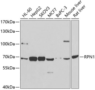
HL-60, HepG2, SKOV3, MCF7, BxPC-3, Mouse liver, Rat liver
Cellular Localization:
Endoplasmic Reticulum Membrane, Melanosome, Single-Pass Type I Membrane Protein.
Calculated MW:
69kDa
Observed MW:
68kDa
This gene encodes a type I integral membrane protein found only in the rough endoplasmic reticulum. The encoded protein is part of an N-oligosaccharyl transferase complex that links high mannose oligosaccharides to asparagine residues found in the Asn-X-Ser/Thr consensus motif of nascent polypeptide chains. This protein forms part of the regulatory subunit of the 26S proteasome and may mediate binding of ubiquitin-like domains to this proteasome.
Purification Method
Affinity purification
Gene ID
6184
RRID
AB_2759339
Buffer Information
Store at -20℃. Avoid freeze / thaw cycles. Buffer: PBS containing 50% glycerol, preserved with proclin300 or sodium azide, pH 7.3.
Western blot analysis of various lysates using RPN1 Rabbit pAb (CAB12497) at 1:1000 dilution. Secondary antibody: HRP-conjugated Goat anti-Rabbit IgG (H+L) (CABS014) at 1:10000 dilution. Lysates/proteins: 25μg per lane. Blocking buffer: 3% nonfat dry milk in TBST. Detection: ECL Basic Kit (AbGn00020). Exposure time: 90s.
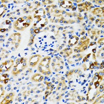
Immunohistochemistry analysis of paraffin-embedded Rat kidney using RPN1 Rabbit pAb (CAB12497) at dilution of 1:100 (40x lens). Microwave antigen retrieval performed with 0.01M PBS Buffer (pH 7.2) prior to IHC staining.
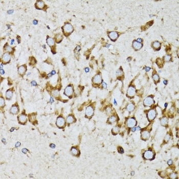
Immunohistochemistry analysis of paraffin-embedded Mouse brain using RPN1 Rabbit pAb (CAB12497) at dilution of 1:100 (40x lens). Microwave antigen retrieval performed with 0.01M PBS Buffer (pH 7.2) prior to IHC staining.